Del Mar Photonics -
Newsletter
Featured inquiry:
Actively stabilized, single frequency ring dye laser with wavelength readout
• Must furnish all optical and mechanical elements for pumping by a vertically
polarized Millenia Pro 5S DPSS laser (532 nm, 5W)
• Output power 800 mW or better at R6G maximum
• Optics set to cover wavelength range 550-650 nm
• Line width of 500 kHz rms/100 ms (or better)
• Mode-hop free scan range of 40 GHz or greater
• Wavelength measurement range at least 400-800 nm with absolute accuracy ± 60
MHz
• Software for wavelength readout ( for Windows XP PC)
• Must provide electronic handshaking signals or software (for Windows XP PC)
for laser scan control
 |
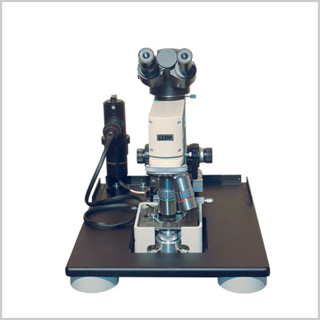
Near Field Scanning Optical Microscope
NSOM Godwit - best spatial optical resolution using the near field scanning optical microscope
(NSOM) principle
Near field scanning optical microscope (NSOM) and atomic force microscope (AFM)
modes of operation
NSOM images with laser and lamp illumination
Commerciaand custom NSOM probes
Near field optica and luminescence images in photon counting mode
NSOM images in collection and illumination modes
Transmission and reflection NSOM configurations
20 nm optical resolution (Raleigh criteria for spatial resolution)
State-of-the-art optical microscope console: simultaneous sample and tip
observation with long working distance objectives
Femtosecond and UV excitation
True single molecule detection
High-resolution AFM imaging of DNA
Godwit-uScope data acquisition and Godwit-FemtoScan image processing software
Ambient light protection with light-tight box
|
 |
New laser spectrometer
T&D-scan for research that
demands high resolution and high spectral
density in UV-VIS-NIR spectral domains - now available with
new pump option!
The
T&D-scan
includes
a CW ultra-wide-tunable narrow-line laser, high-precision wavelength meter,
an electronic control unit driven through USB interface as well as a
software package. Novel advanced design of the fundamental laser component
implements efficient intra-cavity frequency doubling as well as provides a
state-of-the-art combined ultra-wide-tunable Ti:Sapphire & Dye laser
capable of covering together a
super-broad spectral range between 275 and 1100 nm. Wavelength
selection components as well as the position of the non-linear crystal are
precisely tuned by a closed-loop control
system, which incorporates highly accurate wavelength meter.Reserve a
spot in our
CW lasers training
workshop in San Diego, California. Come to
learn how to build a
CW
Ti:Sapphire laser from a kit
|
 |
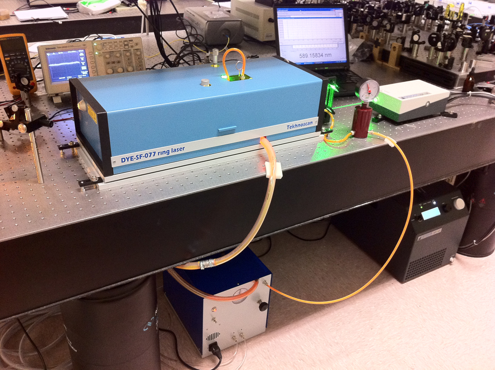
Frequency-stabilized CW
single-frequency ring Dye laser DYE-SF-077 pumped by DPSS DMPLH laser installed
in the brand new group of Dr. Dajun Wang at the The Chinese University of Hong
Kong.
DYE-SF-077 features exceptionally narrow generation line width, which
amounts to less than 100 kHz. DYE-SF-077 sets new standard for generation
line width of commercial lasers. Prior to this model, the narrowest line-width
of commercial dye lasers was as broad as 500 kHz - 1 MHz. It is necessary to
note that the 100-kHz line-width is achieved in DYE-SF-077 without the use of an
acousto-optical modulator, which, as a rule, complicates the design and
introduces additional losses. A specially designed ultra-fast PZT is used for
efficient suppression of radiation frequency fluctuations in a broad frequency
range. DYE-SF-077 will be used in resaerch of Ultracold polar molecules,
Bose-Einstein condensate and quantum degenerate Fermi gas and High resolution
spectroscopy
630nm |
-------------------
Located at the crossroads of biophysical chemistry, optics, nanoengineering, and
ultrasensitive biomedical analysis, research in the
Woehl Lab is
interdiscplinary by nature. It focuses on the optical spectroscopy and imaging
of single molecules and their controlled manipulation using a novel device
pioneered in our research group: the corral trap. The particular aim of our
research is to better understand how molecules use their innate electric fields
to communicate with each other, and how, in turn, we can make use of electric
fields to trap and manipulate single molecules.
1. In many cases, biological molecules carry out their tasks by making use of
molecular electric fields (such as in photosynthesis, enzymatic activity,
electrostatic steering of ligands to active sites, and protein folding and
assembly). In a collaborative effort with the Geissinger group, we employ single
molecules as nanoscale reporters of molecular electric fields at the active
sites of the oxygen carriers myoglobin (see image to the right) and hemoglobin,
where molecular fields may play an important role for the proteins' biological
function. The spectroscopic measurements are carried out at cryogenic
temperatures by applying an external electric field, which shifts the single
molecule absorption or emission line shifts (Stark shift). We have developed two
novel methods of analysis based on quantum chemical calculations that allow for
the determination of internal electric fields (magnitude and orientation) from
the experimental data.
2. All known physical and chemical properties of a molecular species are the
direct result of quasi-electrostatic interactions, and it appears sensible to
focus on electrostatic forces as the most efficient means of exercising control
over single molecules. Based on this idea, we have conceived and developed a new
approach for the trapping and manipulation of single nanoparticles (including
single molecules): the corral trap. Corral trapping of single molecules opens up
new possibilities for the planned (as opposed to purely heuristic) assembly of
molecular-scale devices, detection of single base substitutions (single
nucleotide polymorphisms, SNPs) in the human genome at the single molecule
level, and may lead to new approaches for water purification.
A common component of all our experiments is the ability to image single
molecules with high spatial resolution, and the development and improvement of
instrumentation and theoretical tools for optical microscopy is therefore at the
core of our efforts. In order to spatially localize and select a given molecule,
we utilize widefield microscopy, confocal scanning laser microscopy, or
near-field scanning optical microscopy (NSOM). In aperture-type NSOM, a tapered
optical fiber serves as a nanoscale light source, and we have developed new NSOM
tips on the basis of photonic crystal fibers (see SEM image to the right) with
improved imaging capabilities. We have also mapped the three-dimensional point
spread function (PSF) of a confocal microscope at high spatial resolution and
developed an rigorous theoretical model based on a vectorial approach that has
resulted in PSF Lab, a software package for calculating PSFs that is currently
used by research groups from more than 50 countries worldwide.
Students in my group gain a broad experience in physical chemistry and acquire
specialized skills in a variety of fields such as single molecule imaging,
nanophotonics, laser spectroscopy, thin-film deposition, cryogenic techniques,
metal evaporation, and scanning probe microscopies. Please do not hesitate to
contact me if you wish to participate in any of these research projects.
 |
We are looking forward to hear from you and help you with
your optical and crystal components requirements. Need time to think about
it?
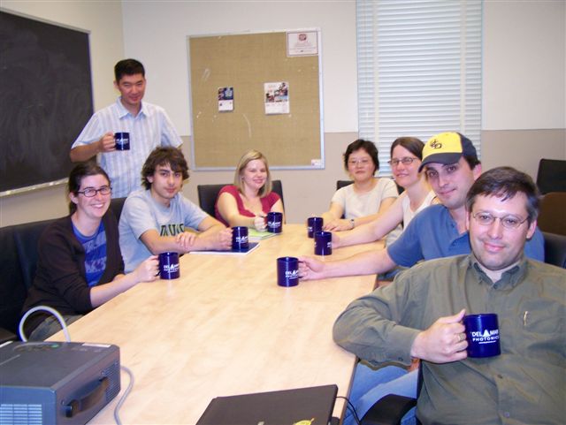
Drop us a line and we'll send you beautiful Del Mar Photonics mug (or
two) so you can have a tea party with your colleagues and discuss your
potential needs. |

Del Mar Photonics, Inc.
4119 Twilight Ridge
San Diego, CA 92130
tel: (858) 876-3133
fax: (858) 630-2376
Skype: delmarphotonics
sales@dmphotonics.com


