Featured publications abot research with
NSOM Godwit
Two-Photon Absorption Near Field Imaging of Non-Fluorescent Organic
Nanoparticles
Jeffery E. Raymond, Theodore Goodson III*
Departments of Chemistry and Macromolecular Science & Engineering, 930 N.
University Ave., University of Michigan, Ann Arbor MI 48109
We present here the first reported use of fiber aperture near-field optical
microscopy (NSOM) for the purpose of characterizing directly the two-photon
absorption (TPA) of non-emissive nanoparticles. It will be displayed how this
empirically driven technique can provide per particle and per molecule
assessment of the two-photon cross-section (TPACS) by extracting the non-linear
optical (NLO) signal from that due to scattering and far-field effects. This is
shown with the investigation of a known two-photon responsive porphyrin dimer,
which has exhibited both severe fluorescence quenching and a multiple order of
magnitude TPACS enhancement in aggregate, after self-assembly into uniform
nanoparticles. A particular emphasis will be placed on the viability of this
technique for the characterization of low-scatter optical limiting organic
nanomaterials.
NSOM, two-photon, porphyrin dimer, organic nanoparticle
Single-Particle Two-Photon Absorption Imaging and Enhancement Determination for
Organic Nanoparticles
Jeffery E. Raymond and Theodore Goodson, III*
Departments of Chemistry and Macromolecular Science & Engineering, University of
Michigan, 930 North University Avenue,
Ann Arbor, Michigan 48109, United States
The lack of characterization regimes available for the rapid single particle
assessment of two-photon (TPA) response in nanomaterials remains a critical
barrier to nonlinear optical device development. This is particularly true of
nonemissive species whose TPA must often be characterized in the bulk. In this
study, self-assembly is used to produce uniform nanoparticles from a novel
porphyrin dimer, which is known to exhibit both severe fluorescence quenching
and two-photon cross section (TPACS) enhancement when assembled into
macromolecules.
We present here the first reported use of fiber aperture near-field optical
microscopy (NSOM) for the purpose of characterizing directly the TPA of
nonemissive nanoparticles, observing directly a 5-fold enhancement in TPA
response. This assembly/characterization regime provides a fast and fully
actualized method for the generation of low-scatter optical-limiting organic
nanomaterials where domain size, morphology control, and TPA enhancement are all
critical to application viability and unobservable via bulk measurements.
Quantitative Non-Linear Optical Imaging in the Nano- Regime
Jeffery Raymond, Theodore Goodson III
Department of Chemistry, Department of Macromolecular Science and Engineering
University of Michigan, Ann Arbor Michigan
The development of functional solid state non-linear optical (NLO) systems for
device applications is critical to several fields. Optical computing, laser
hardening, 3-dimensional data storage and remote sensing are just a few of the
areas advanced by the characterization of new NLO systems. One promising venue
for the development of these technologies is the nano-/meso-scale self assembly
of viable chromophores into tunable aggregates. Here we present a method by
which individual aggregates can be quantitatively imaged by two photon
fluorescence near field scanning optical microscopy (NSOM).
two-photon, near-field, TPEF, TPA, NSOM, rhodamine B
Two-Photon Enhancement in Organic Nanorods†
Jeffery E. Raymond,‡ Guda Ramakrishna,‡ Robert J. Twieg,§ and Theodore Goodson
III*,‡
Department of Chemistry, Macromolecular Science and Engineering, Gerald R. Ford
School of Public Policy,
UniVersity of Michigan, Ann Arbor, Michigan 48109, and Department of Chemistry,
Kent State UniVersity, Kent, Ohio 44242
The nonlinear optical response in one-dimensional organic nanorods of
N,N-dimethyl-4-4((4-(trifluoromethylsulfonyl)phenyl)ethynyl)aniline (DMFSPA) was
investigated to probe the long-range interactions in the nanocrystals on the
microscopic level. Differences in the linear and nonlinear optical properties
are shown for two different morphologies of these organic crystals as well as
for the chromophore in solution. The optimized nanocrystalline suspension had
more than an order of magnitude increase in the two-photon excited fluorescence
when compared to the solution phase of DMFSPA at similar chromophore densities.
The one and two-photon properties of the nanocrystals and bulk crystals are
compared by near-field scanning optical multiscope imaging. The images also
provide insights into the formation of the nanorods during initial
crystallization, changes in the optical response of the system with time, and
the viability of these and similar nanomaterials for consideration in
solid-state organic device applications. In addition to providing an imaging
regime by which to assess this and other solid-state nanocrystalline organics,
our investigation provides a simple and elegant method for enhancing the
nonlinear optical response of organic materials by transition to nanoscale
morphologies, without the need for additional chemical modification or
synthesis.
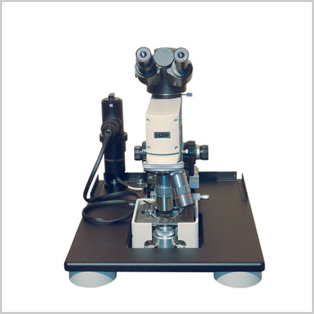
Near Field Scanning Optical Microscope NSOM Godwit -
request a quote
Near Field Scanning Optical Microscope NSOM Godwit is a device that enables you to get the best spatial optical resolution using the near field scanning optical microscope
(NSOM) principle
Near field scanning optical microscope (NSOM) and atomic force microscope (AFM)
modes of operation
NSOM images with laser and lamp illumination
Commerciaand custom NSOM probes
Near field optica and luminescence images in photon counting mode
NSOM images in collection and illumination modes
Transmission and reflection NSOM configurations
20 nm optical resolution (Raleigh criteria for spatial resolution)
State-of-the-art optical microscope console: simultaneous sample and tip
observation with long working distance objectives
Femtosecond and UV excitation
True single molecule detection
Godwit-uScope data acquisition and Godwit-FemtoScan image processing software
Ambient light protection with light-tight box
Fields of application:
Molecular spectroscopy, Fluorescence, Surface science, Thin films, Biology,
Chemistry, Solid state physics, Nanotechnology, Material Science, Medicine,
Education, Fiber optics
Specifications
XY sample scanner
20 mm diameter central opening
Maximum scan size: 40 μm x 40 μm
Minimum scan step: 0.01 nm
Maximum image size: 1024 x 1024 pixels
Maximum XY sample travel: 10 mm x 10 mm (computer controlled)
Optical resolution
50 nm typical (depends on NSOM probe and sample under investigation)
Piezo-inertial Z stage with mounted NSOM probe
Maximum Z-travel: 9 mm
Z-scanning range: ±5 μm
Electronic control unit
XY stage electronics
Z stage electronics
Feedback (shear-force) electronics
Photon counting electronics
Lock-in amplifier
Connected to a computer via PCI card
Photon counter
Maximum photon counting rate: 5 x 10⁷ cps
Dark counts: < 10 cps
Spectral response: 185 - 680 nm (185 nm - 850 nm optional)
Standard illumination sources
150 W quartz - halogen lamp
670 nm laser diode
532 nm solid state laser
488 nm Ar-ion or solid state laser
400 nm femtosecond laser
Recommended NSOM probes
633 nm single mode, Al - coated (50 - 80 nm aperture), 100 kHz resonance
400 nm single mode, Al - coated (<100 nm aperture), 32 kHz resonance
Dimensions
Optical unit with light-tight box: 350 (W) x 460 (D) x 460 (H)
Electronic control unit: 19” rack mountable or 170 (W) x 420 (D) x 200 (H)
request a quote - request a brochure - request a manual
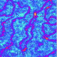
AFM (topography) image of DNA (<3 nm thickness), |
Del Mar Photonics nano-imaging gallery
High resolution MFM image of Seagate Barracuda 750Gb Hard Drive, ST3750640AS.
130 nm Ag nanoparticles immobilized on the metal surface, 3.6x3.6 um scan
Magnetic structure of surface domains in Yttrium Iron Garnet (YIG) film
Atomic resolution on HOPG obtained with the 100 micron scanner
NSOM Fluorescence image of 100 nm - diameter TransFluoSpheres
Near-field optical image of 250 nm - diameter gold beads, deposited onto a glass
slide
AFM (topography) image of DNA (<3 nm thickness),
deposited onto a glass slide
Near-field optical image of 100 nm - diameter polystyrene beads, deposited onto
a glass slide