Del Mar Photonics -
Newsletter
6th Biomarker Development – March 15-16, 2012 – San Diego, CA
Day 1 - Thursday, March 15, 2012
7:00a Registration & Continental Breakfast
7:55a Welcome & Opening Remarks
Plenary Session
8:00a Regulatory Update: Drug Development Tool
Qualification at the FDA and EMA – What It Is and
What It Isn’t
Carolyn Compton, MD, Ph.D.
President & CEO
Critical Path Institute
Both regulatory agencies and the medical product
industry recognize opportunities for new biomarkers and
methods to get effective, safe medicines to patients
faster. However, developing consensus on the utility of
new biomarkers and establishing a process for putting
them into routine practice for regulatory decision making
in medical product development is a complex
undertaking. In 2009 and 2010, respectively, the
European Medicines Agency and the United States Food
and Drug Administration released guidance that
describes a voluntary pathway for “qualification” of novel
drug development tools or methods. Determining the
relative advantages/disadvantages and evidentiary
standards required for regulatory “qualification,” applying
a novel biomarker on a specific drug development
program, or submitting an application for a diagnostic
device are challenging tasks.
The Critical Path Institute leads five pre-competitive
consortia that develop and qualify safety, imaging, and
disease biomarkers as well as patient-reported outcomes
instruments. Several consortia use Clinical Data
Interchange Standards Consortium disease area data
standards in constructing clinical trial databases from
which disease progression models are developed.
Specific examples from these consortia will be utilized to
illustrate the distinct, but sometimes overlapping,
regulatory pathways available for reducing uncertainty in
the use of new medical product development tools.
• Understand the drug development tool qualification
process at FDA and EMA
• Learn how pre-competitive consortia and data-sharing
are driving adoption of novel drug development tools
• Appreciate the advantages and challenges of the
various regulatory pathways for utilizing novel
biomarkers in drug development
8:30a Keynote Speaker
Steven J. Skates, Ph.D.
Associate Professor of Medicine
Massachusetts General Hospital Cancer Center
Keynote Speaker
9:00a Grants for Targets and Biomarkers – A Novel
Open Innovation Initiative in Drug Discovery
Khusru Asadullah
Vice President, Head of Global Biomarker
Bayer HealthCare
Collaborations between industry and academia are
steadily gaining importance. To combine expertises Bayer
Healthcare has set up a novel open innovation approach
called Grants4Targets. Ideas on novel drug targets and
biomarkers can easily be submitted on
www.grants4targets.com. After a review process, grants
are provided to perform focused experiments to further
validate the proposed targets/biomarkers. In addition to
financial support specific know-how on target validation
and drug discovery is provided. Experienced scientists are
nominated as project partners and, depending on the
project, tools or specific models are provided. Around 600
applications have been received and 77 projects granted.
According to our experience, this type of bridging fund
provides a valuable tool to foster drug discovery
collaborations.
9:30a Networking and Refreshment Break
Personalized Medicine for Non-Oncology Uses
(Dual Track)
10:00a Revealing Rheumatoid Arthritis at the
Molecular Level- Build It and They Will Come?
Lucy Lu, Vice President Business Development,
Crescendo Bioscience
Over 1M people in the North America live with rheumatoid
arthritis (RA)- a chronic autoimmune disease that affects
joint function and often leads to disability. Although there
are several conventional DMARDS and nine targeted
biologic drugs approved for treating RA, there is no
treatment guideline or lab test to guide therapy selection.
“Treating to Target“ has emerged as an important principle
for the management of RA in the last several years.
Various metrics, usually combine swollen/tender joint
counts, physician assessment and patient questionnaire,
6th Biomarker Development – March 15-16, 2012 – San Diego, CA
have been used to monitor disease activity and guide
practice to help RA patients reach the desired “target”
state.
This talk will describe the development and
commercialization of a novel multi-biomarker RA disease
activity test, Vectra™ DA, which is aimed at providing an
objective and quantitative measurement to improve RA
patient management.
• What‟s the science behind the test?
• How has the test been helping rheumatologists manage
their RA patients?
• What are the barriers and challenges of
commercializing a novel Dx test?
• Is the test useful in new RA drug development?
10:30a Biomarkers to Predict the Response to
Rituximab
Cornelis L. Verweij, Ph.D., Head Inflammatory Disease
Profiling, VU University Medical Center
Rituximab (Mabthera®/Rituxan®) is a therapeutic
monoclonal antibody against CD20 expressed on B cells,
which is effective in depleting B cells and shown to be
highly beneficial in decreasing clinical symptoms, safe
and well tolerated in the treatment of rheumatoid arthritis.
However, clinical experience revealed that not all
patients do respond to treatment. Since nearly all treated
patients experience an effective depletion of circulating B
cells, questions have been raised concerning the
mechanism of action. This presentation provides
strategies and outcome of biomarker discovery research
to predict the response to B-cell depletion therapy using
rituximab. The results add new and important information
to our understanding on the mechanism that underlies
the clinical outcome of rituximab treatment and
demonstrate clinical utility for the use of a specific set of
genes based on Receiver Operating Characteristic
(ROC)-curve analysis (AUC 0.87) to predict nonresponse
to rituximab in RA. This biomarker set is likely
to become a substantial aid to the physician, taking the
paradigm of personalized medicine one step further.
Benefits:
• Understand the unmet clinical needs in targeted
therapies with biologicals
• Understand approaches for biomarker discovery
• Understand the pharmacology of B-cell depletion
associated with clinical response
• New insight in the prediction of response outcome to Bcell
depletion
11:00a Biomarkers And Personalized Medicine: The
Time Is Now
Claudio Carini, M.D., Ph.D., FRCPath, Translational
Medicine BioTx Clin Res, Pfizer
Next Generation Sequencing and Discovery of
New Diagnostics (Dual Track)
11:30a Daniel R. Salomon, M.D., Associate professor,
Department of Molecular and Experimental Medicine,
The Scripps Research Institute
12:00p Luncheon on Your Own
Biomarker Validation of Preclinical to Clinical
Studies
1:30p Discovery and Development of Biomarkers for
Clinical Trials Using Preclinical Animal Studies
Stanley Belkowski, Principal Research Scientist, Target
Validation Team, Janssen Research and Development
At Janssen Immunology, the biomarker team is
responsible for identifying and implementing appropriate
pharmacodynamic, mechanism of action and personalized
medicine measurements into all phases of clinical
development. Informative biomarkers are enablers of
decision making and are utilized during early clinical trials
to modify Phase I protocols and to accelerate design of
future trials. In this presentation, I describe the
identification, development and implementation of
biomarkers for an early stage asset moving into Phase I
trials. Examples of how critical path biomarkers were
developed to help make decisions in disease indication
and target engagement will be illustrated. In this program,
we employed in vitro and animal studies to develop key
biomarkers for implementation in upcoming clinical trials to
make decisions on dose ranging and efficacy in disease.
These data will provide the compound development team
with increased insight into the molecular effects of the test
agent and facilitate early decision making for the next
stages of development.
This talk will:
• Outline paths of biomarker discovery and development
from preclinical to proposed clinical use
• Examine both gene signature and protein markers
• Explore alternative sources of biomarker samples
• Show the beneficial use of in vitro and preclinical animal
samples for biomarker development
6th Biomarker Development – March 15-16, 2012 – San Diego, CA
1:55p Translation of Clinical Biomarkers to
Preclinical Models in Cardiovascular Diseases
Shian-Jiun Shih, Head, Molecular Biomarkers,
Translational Medicine Research Centre, MSD
2:20p Statistical Methods for Identifying
Biomarkers to Reduce Disappointment in the
Validation
Timothy R. Church, Professor, Department of
Environmental Health Sciences, School of Public Health,
University of Minnesota
Background: Candidate biomarkers for early detection
often fail on validation. The causes are introduced during
the initial development process. Minimizing their
influence can reduce the disappointment at validation.
Although the process of identifying biomarkers for early
detection can vary widely, it is possible to embed the
development process in an algorithm that allows the
researcher to correct the apparent accuracy of the
biomarker for biases that creep in. This applies to
combinations of markers as well as to single markers.
Methods: Early detection is prediction. To sift through a
set of biomarkers for single-marker or combined-marker
candidates, some process is applied to measure the
quality of each candidates predictive accuracy in a
development sample and select the best one. The
measure might be the maximum area under a receiver
operating characteristic curve or sensitivity and
specificity. The process of selecting these candidates
determines how big the net bias will be. To ensure that
this bias can be estimated and corrected for the
investigator must account for all steps in the
development. Using a bootstrap resampling method that
applies the entire development process to each
bootstrap sample permits the estimation of bias within
the development sample and the projection of the
estimated accuracy to a validation sample.
Results: Bias depends upon the development method
and the sample size. Bootstrap resampling, appropriately
done, identifies the bias so it can be corrected, yielding
more realistic estimates of accuracy.
Conclusions: Carefully planned development processes
coupled with bootstrap cross-validation can reduce
disappointment in the validation step of biomarker
research.
Benefits:
• Understanding the process by which bias is introduced.
• Motivating rigor in the process of selecting candidate
biomarkers.
• Flexibility in choosing measures of predictive accuracy.
• Providing a workable tool to reduce bias in estimates of
accuracy.
2:45p Networking & Refreshment Break
3:15p Discovery of Predictive Biomarkers for Patient
Selection in Oncology Trials Using Pre-clinical Models
of Cancer
Adam Pavlicek, Ph.D., Computational Biology, Oncology
Research Unit, Pfizer
Identification of predictive biomarkers is essential for the
success of targeted cancer therapies. I will review our
current approach to biomarker discovery and precision
medicine in oncology. Specific cases of CDK4/6 and
gamma-secretase small molecule inhibitors will be
presented. Key computational and statistical aspects of
predictive biomarker studies will be mentioned.
Key benefits:
• Identification of predictive biomarkers in pre-clinical is
essential for early implementation of patient selection
strategies in clinical trials
• Specific cases of CDK4/6 and gamma-secretase
inhibitors will be presented
• Different therapeutic targets may require different preclinical
models
• Rigorous analytical and statistical framework needs to
be developed to limit false discoveries
Shortening Development times with Biomarker
Defined Patient Populations
3:45p Accelerating In Vitro and In Vivo Biomarker
Research: From Biomarker Discovery to Late Stage
Clinical Projects
Yoshi Oda, President, Biomarkers & Personalized
Medicine, Eisai
Biomarkers enable early confirmation of proof-ofmechanism
(POM) and efficacy of drug candidates, and
stratification of patients who may benefit from drug
candidates. Therefore biomarkers can enhance the speed
and accuracy of the overall process of drug development.
The timing of biomarker research is critical to shorten
timeline of drug development. The achievement of
preclinical POM in animal models followed by the clinical
POM can be the first decision point of the project. In
6th Biomarker Development – March 15-16, 2012 – San Diego, CA
general, a pharmacodynamic (PD)marker should be
linked to POM and the PD marker can be used to
determine the right dose of the drug candidate in earlier
clinical study. The development of assay systems for
POM and PD markers is essential during animal model
studies. Patient stratification biomarkers are also
important to identify the right patient population, which
can increase the success rate of clinical trials and reduce
the size of patient recruitment for the study. Patient
stratification biomarkers should preferably be the direct
target of the drug candidates, however other molecules
up or down stream of the target may be suitable
biomarkers. Those indirect molecules for patient
stratification could be identified by retrospective analysis
of clinical samples. This is probably the most real
situation, but we need to identify those biomarkers during
preclinical studies to shorten timeline of clinical study.
Our unit has been organized to conduct from biomarker
discovery to clinical validation. We have multiple-omics
technologies, IHC/FISH, ELISA, qPCR, preclinical
imaging and bioinformatics. I present current our
challenges.
4:15p [Oral Presentations from Exemplary
Submitted Abstracts]
To be considered for an oral presentation, please submit
an abstract here
5:00p Networking Reception
Day 2 - Friday, March 16, 2012
Biomarkers in Clinical Trials
8:00a A Mouse Model of Doxorubicin-Induced
Chronic Cardiotoxicity to Identify Predictive
Biomarkers of Cardiac Tissue Injury
Varsha G. Desai, Ph.D., Research Biologist, Center for
Functional Genomics, Division of Systems Biology, U.S.
FDA/National Center for Toxicological Research
Doxorubicin (DOX) is a potent chemotherapeutic drug
known to cause dose-related cumulative and irreversible
cardiomyopathy in cancer patients. Currently, cardiac
troponins are considered clinical biomarkers of cardiac
injury in cancer patients. However, troponins identify
cardiac toxicity only after tissue damage has occurred. In
our laboratory, we have developed a mouse model of
DOX-induced chronic cardiotoxicity to aid in the
development of predictive biomarkers of cardiotoxicity.
Male B6C3F1 mice were administered a weekly dose of
3 mg/kg DOX, or an equivalent volume of saline,
intravenously via tail vein for 4, 6, 8, 10, 12, and 14
weeks, resulting in cumulative DOX doses of 12, 18, 24,
30, 36, and 42 mg/kg, respectively. Results indicated a
significant increase in plasma cardiac troponin T levels in
all mice exposed to cumulative DOX doses of 12 mg/kg
and higher compared to saline-treated controls, indicating
cardiac tissue injury. In addition, a dose-related increase
in severity of cardiac lesions was noted in mice following
cumulative DOX exposures to 24 mg/kg and higher.
Preliminary analysis of hearts by transmission electron
microscopy revealed mitochondrial swelling with
disorganization, fragmentation and loss of cristae at 12
mg/kg DOX. Altogether, these findings demonstrate the
development of a mouse model of DOX-induced chronic
cardiotoxicity.
Outline and benefits:
• Development of a mouse model of DOX-induced
chronic cardiotoxicity
• Mitochondrial damage occurs prior to cardiac lesions
during DOX exposure
• Aid in the identification of novel predictive biomarkers of
DOX-induced cardiotoxicity
• Translation of predictive biomarkers to the clinic would
be useful in designing optimal dosing regimen
8:45a Progression of Biomarkers for Alzheimer'
Disease Diagnosis
Johan Luthman, Senior Program Leader, Early
Development, Neuroscience & Opthamology R & D,
Merck
9:10a Epigenetic Immune Cell Markers – Robust
Results from Frozen Whole Blood and FFPE Tissue
Samples in Clinical Trials
Ulrich Hoffmueller, Chief Business Officer and Founder,
Epiontis
The responsiveness of the immune system to counter
unwanted antigens is a prominent medical parameter.
This responsiveness is determined by the cell counts and
ratios of different leukocyte populations, which are
promising diagnostic approaches. Current standard
methods - FACS in peripheral blood,
Immunohistochemistry (IHC) in solid tissue - have a
limited ability for exact cell quantification owing to
arbitrary/subjective gating protocols for FACS and lacking
precision for IHC. For regulatory T-cells (Tregs), additional
problems are manifested due to lack of specificity of Treg
identifying proteins (CD25, FoxP3). Both proteins are
synthesized also in activated effector T-cells, leading to
detection of mixed cell populations.
The discovery of cell type specific epigenetic markers
allows precise quantitation of immune cells in all human
6th Biomarker Development – March 15-16, 2012 – San Diego, CA
samples. The tests are based on quantitative PCR
targeting genomic DNA. Therefore, readout is stable and
samples can be frozen and easily shipped. This allows
monitoring of patients in multicenter studies,
retrospective studies, comparison of results between
different studies, and routine monitoring.
In addition to technical benefits, higher Treg specificity is
observed using epigenetic FOXP3 gene analysis,
showing epigenetic activation only in true Tregs and
epigenetic suppression in all other tested cells.
Application of epigenetic assays for Tregs, overall T-cell
population, NK cells and further subpopulations will be
presented in cancer and immune disease patients. The
results reveal the same trends as measurements with
alternative methods, while showing higher precision.
Furthermore, the tests can be applied on both blood and
tissue allowing measurements of circulating and tissueinfiltrating
immune cells.
Analytics in Biomarker Development
9:35a A few Genome-Wide Scores Allow Predictive
and Stable Tumor Characterization
Andreas Schuppert, Director, Bayer Technology
Services
The identification of reliable models for the prediction of
abiotic stress response and disease propagation is
crucial for a wide range of challenges for biomarker
development. Improved, personalized therapies and
reduction of attrition rates in drug development are
examples for applications which will strongly benefit from
further progress in model development.
Because the detection of prognosis-related signals in
heterogeneous clinical data is strongly hampered by
biological heterogeneity, we aimed to develop an
approach allowing the deconvolution of the respective
patterns in the data.
On the example of a metaanalysis of genome-wide
expression studies for solid cancers, the patterns which
are associated to tumor classification will be discussed
with respect to their potential for clinical biomarkers. We
show that genome-wide analysis allows the
deconvolution of patterns in biological data according to
impacts from environmental, primary and secondary
response on clinical phenotypes and drug efficacy.
Moreover, also the accuracy of biomarkers based on
single genes strongly depends on specific genome-wide
expression patterns. We provide a quantitative mean to
assess genome-wide expression studies with respect to
their information content, accuracy and stability of
biomarkers.
• Predictive assessment of cross-study stability of
expression based biomarkers
• Predictive assessment of accuracy of biomarkers
based on genome-wide expression data
• Mapping of genome wide expression patterns onto a
few expression scores
• Stable tumor characterization using three genome-wide
scores.
10:00a Networking & Refreshment Break
10:30a The Automated Micro-fluidic Immunoassay
(AMI) for Superior Protein Analytics
Urban Kiernan, Senior Scientist, Thermo Fisher
Scientific
Since the establishment of protein based clinical analytics
some four decades ago, the science in this field has been
ever evolving. Improvements regarding speed, efficiency
and analytical sensitivity have dominated the
developmental landscape; however, the current standard
is still based on utilizing micro-titer wells as the reaction
sites. Even though this format provides the user with many
desirable attributes, such as high throughput analysis, it is
several decades old and is the contributing factor of the
rate limitations of conventional immunoassays.
Described is a novel application that converts the classical
immuno-assay approach from the micro-titer plate into a
dynamic micro-fluidic format , which retains the high
throughput capacity and enhances both the speed and
dynamic range of the assay. Termed the Automated
Micro-fluidic Immunoassay (AMI), all the steps utilized in a
classical ELISA format are translated into 3D micro-fluidic
structures that are outfitted in the opening of a functional
pipette tip. All incubation and reaction steps are performed
in these micro-fluidic devices with the final reacted
substrate being deposited into a standard micro-titer plate
for non-subjective UV/Vis absorbance measurement. This
hybrid approach allows for the user to capitalize on all the
known benefits of micro-fluidic applications, but maintains
the attributes that make the dominance of the micro-titer
plate based assays (i.e., high throughput, non-subject
detection, etc.
This presentation will describe the AMI approach in full
detail and provide examples that run head-to-head with
current ELISA. Data will be provided from model systems
that show the reproducibility and accuracy of the AMI and
demonstrate clear improvements that will enhance
productivity and ameliorate many pinch points that are
commonly observed in both clinical and research studies.
Benefits of the Talk:
• Explain the advantages of the AMI using micro fluidic
6th Biomarker Development – March 15-16, 2012 – San Diego, CA
immuno-affinity devices (in a pipette tip format) for
enhanced enrichment and accelerated reaction times.
• Demonstrate the use of the AMI micro immuno-affinity
pipette technology with classical color-metric detection
methods.
• Describe the superior analytical characteristics of AMI
compared to comparable well based approaches.
• Describe the pinch points in current analytical work
flows which would most benefit from the integration of
the AMI technology and process.
New Technologies & Novel Approaches in
Biomarker Development
10:55a Karyo-instability and Biomarker Expressions
in Tumors
Leon Bignold, Associate Senior Lecturer, University of
Adelaide
Accumulations of both nucleotide sequence errors in
DNA and mitotic and chromosomal abnormalities (karyoinstabilities)
are characteristic of malignant tumors. In the
same tumors there are usually reductions, but also
occasional supra-normal biomarker expressions. If both
the sequence errors and the karyo-instability cause lossof-
function mutations, these supra-normal expressions
should not occur.
HJ Muller (1) suggested that in species of organisms
which accumulate mutations, extinction is avoided by
crossing-over of chromosomes during meiosis. This
phenomenon allows good copies of viability-essential
genes to be exchanged for mutant copies in some
gametes, which lead to new non-mutant individuals. The
present author (2) has suggested that in the immortality
of tumor cell populations, unequal distributions of
chromosomes in anaphase play a similar role. These
“asymmetric mitoses” may create the immortal tumor
cells by delivering to them extra good copies of viabilityessential
genes.
This scenario in mitosis of tumor cells indicates possible
value of karyo-stabilising agents: (i) in reducing growth
rates of karyo-unstable cancer cells, (ii) in reducing
incidence of tumors appearing in „at risk‟ tissues, such as
the bronchial epithelium in ex-smokers, and/or the early
(pre-blast transformation) phase of chronic myeloid
leukemia.
Summary:
• Possible solution to a paradox associated with genomic
instabilities
• Explanation of common biomarker expression findings
• Suggestion: any agent which could stabilize mitoses and
chromosomes might be of value to patients with cancers,
and also possibly to individuals with „at risk‟ tissues.
[1] Muller HJ. Mutat Res 1964 1: 2-9.
[2] Bignold LP. Cancer Genet Cytogenet, 2007 178: 173-
4.
11:20a Beyond HDL Cholesterol: Application of MRM
Proteomics to the HDL Proteome
Hussein Yassine, MD, Director, Lipid Clinic; Assistant
Professor, Division of Endocrinology, University of
Arizona
In this talk, I will explore the application of Multiple
Reaction Monitoring (MRM) on cardiovascular disease,
and understanding high density lipoprotein structure (HDL)
and function. Recent proteomic studies indicate that HDL
consists of a lipid core and a wide array of attached
proteins involved in inflammation and blood clot formation.
Saturated fat intake in the diet impairs the function of HDL
on endothelium by mechanisms not related to current
concepts of HDL cholesterol (HDL-C). We hypothesize
that subjects with diabetes (DM) and cardiovascular
disease (CVD) have HDL enriched with inflammatory and
prothrombotic proteins, and that saturated fat ingestion
illustrates one mechanism for generating inflammatory
HDL particles that are linked to endothelial dysfunction.
We isolated HDL by ultracentrifugation and defined 60
HDL attached proteins using Multiple Protein Identification
Technology “MudPIT”. We then selected representative
tryptic peptides from each protein for Multiple Reaction
Monitoring (MRM) analysis. Using MRM, we investigated
the change in HDL proteins in subjects with and without
diabetes and cardiovascular disease. The talk will
highlight the application of a high throughput technology
into the discovery and verification of biomarkers relevant
to cardiovascular disease, and the lipoproteome in
particular. Multiple Reaction Monitoring (MRM) allows the
simultaneous monitoring of 100s of proteins in very low
sample volumes and with improvement in the costs of
conducting this essay, it will likely replace immunoassays
given the lack of dependence on existing antibodies.
11:45a Luncheon
1:00p Next Generation X-aptamers for Diagnostics
and Therapeutics
6th Biomarker Development – March 15-16, 2012 – San Diego, CA
David Gorenstein, Ph.D., Associate Dean for Research /
Founder AM, The University of Texas Health Science
Center at Houston and AM Biotechnologies
We have developed novel, next-generation modified
DNA oligonucleotide aptamers selected from large
combinatorial libraries to target a number of protein
biomarkers. We have developed both in vitro enzymatic
combinatorial selection and split-synthesis chemical
combinatorial methods to identify phosphorothioatemodified
oligonucleotide “thioaptamers” and next-gen
“X”-aptamers to a number of different protein targets for
both proteomics and biomarker discovery. The Xaptamers
also include a large range of chemical (X)
modifications to the 5-X-dU position and thus represent a
hybrid of aptamer backbone, protein amino acid-like
sidechains, and small molecule leads in a self-folding
scaffold that can be readily identified by oligonucleotide
sequencing. Compared to conventional aptamers, this
approach dramatically expands the chemical diversity
that can be incorporated to select X-aptamers with high
affinity for diverse molecular biomarkers. Large beadbased
combinatorial libraries of these aptamers can be
rapidly selected. These X-aptamers and thioaptamers
are being used as antibody substitutes in biomarker
identification to tumor cells and tumor vasculature and in
various microfluidics and mass spec chips for proteomics
and diagnostics. Examples of application of the beadbased
X-aptamer selection are demonstrated for
targeting cancer tissue and cells expressing CD44.
Development of Next Generation X-aptamers Targeting
Biomarkers
• How are X-aptamers better than antibodies?
• How do we improve on aptamers for increased affinities
and specificities?
• How can we create next-generation aptamers with
amino acid-like sidechains, and small molecule leads
using novel bead-based combinatorial libraries?
• What biomarkers can we target with these
technologies?
• What is the IP landscape for developing these nextgeneration
X-aptamers?
1:25p miRNA’s as Biomarkers – Do current
profiling methodologies support their use?
Philip Z. Brohawn, Associate Scientist, Translational
Sciences, Medimmune
Since their characterization in the early 1990‟s, hundreds
of miRNA‟s encoded in the human genome have been
identified. miRNA‟s are small, less than 22 bp, noncoding
RNA‟s thought to play a major role in the
differential regulation of mRNA targets. Because of this
functionality, miRNA have the potential to act as an
additional source of biomarkers. However, the small
nature of miRNA‟s, as well as the dynamics of their
expression, make reliably and reproducibly assessing the
expression of these biomolecules a challenge by
traditional expression profiling methodologies. To turn
miRNAs into robust biomarkers, tools for reliably and
reproducibly measuring them in blood or tissues are
essential. To this end, we evaluated multiple miRNA
expression profiling platforms utilizing hybridization,
Taqman and Sybr based chemistries to assess their
accuracy, reproducibility, throughput and applicability in
the clinical trial setting. We utilized next generation
sequencing, and more specifically miRNA seq, as a
reference. Conclusions from this study will help guide the
selection of higher throughput cost-effective platforms
when including miRNA expression profiling in a clinical
setting.
• Provides a platform guide for validation of miRNA‟s as
biomarkers
• Provides a practical comparison of multiple miRNA
expression profiling platforms
• Helps to further understanding of current miRNA
expression methods
• Demonstrates power of Next Generation Sequencing
miRNA seq application
• Will benefit investigators who want to evaluate miRNAs
as biomarkers in clinical trials
1:50p Hypothesis Free Biomarker Discovery
Platform Evolving into your Companion Diagnostics
Gitte Pedersen, Chief Executive Officer, Chief Executive
Officer, Genomic Expression Inc
Genomic Expression‟s hypothesis-free biomarkerdiscovery
platform uses intelligent target-filtering
technology integrated with sequencing short reads on a
small chip addressing the sample prep and data analysis
bottlenecks in current sequencing platforms.
Prior to the wet lab, we use bioinformatics to optimize our
applications using existing sequence data. Then we
deploy an intelligent target-filtering process, which
generates a single-stranded tag library. Because the tags
are single stranded, it is possible to detect all possible
tags using a universal high-throughput platform. The cost
per sample is low, the content/test very high and
hypotheses free.
Our approach represents a paradigm shift in the
biomarker-discovery process, saving up to $20 million and
more than two years in pre-clinic testing while increasing
the probability of success.
We are developing genomic signatures that translate into,
for a doctor, easy-to-understand information regarding
treatment options for cancer patients, supported by a $30
million grant project where we have access to 15 million
6th Biomarker Development – March 15-16, 2012 – San Diego, CA
biological samples associated with cradle-to-grave
electronic medical records.
We partner with pharma and diagnostic companies to
provide access to the platform in a service and solution
model.
2:15p Building Multidimensional Biosignatures of
T2D and CVD Based on Protein Microheterogeneity
Randal Nelson, The Biodesign Institute, Arizona State
University
Protein microheterogeneity, as it occurs through healthy
and diseased populations, is a largely unexplored area of
biomarker development. Here, we used targeted mass
spectrometric approaches to view subtle, dynamic
changes in the proteome of healthy individuals and
patients with type 2 diabetes (T2D) and related
cardiovascular diseases (CVD). Molecular heterogeneity
was observed in a number of plasma proteins, and was
grouped into panels to create a multidimensional view of
healthy – to – T2DM – to – CVD that aligns with disease
pathobiologies. Presented are results from viewing T2D
and CVD populations that indicate the potential of
subtyping individuals within the T2D/CVD continuum.
2:40p John Blume, Chief Science Officer, Applied
Proteomics
3:15p Conference Concludes
4th Oncology Biomarkers – March 15-16, 2012 – San Diego, CA
Day 1 - Thursday, March 15, 2012
7:00a Registration & Continental Breakfast
7:55a Welcome & Opening Remarks
Plenary Session
8:00a Regulatory Presentation
Carolyn Compton, MD, Ph.D.
President & CEO
Critical Path Institute
Both regulatory agencies and the medical product
industry recognize opportunities for new biomarkers and
methods to get effective, safe medicines to patients
faster. However, developing consensus on the utility of
new biomarkers and establishing a process for putting
them into routine practice for regulatory decision making
in medical product development is a complex
undertaking. In 2009 and 2010, respectively, the
European Medicines Agency and the United States Food
and Drug Administration released guidance that
describes a voluntary pathway for ―qualification‖ of novel
drug development tools or methods. Determining the
relative advantages/disadvantages and evidentiary
standards required for regulatory ―qualification,‖ applying
a novel biomarker on a specific drug development
program, or submitting an application for a diagnostic
device are challenging tasks.
The Critical Path Institute leads five pre-competitive
consortia that develop and qualify safety, imaging, and
disease biomarkers as well as patient-reported outcomes
instruments. Several consortia use Clinical Data
Interchange Standards Consortium disease area data
standards in constructing clinical trial databases from
which disease progression models are developed.
Specific examples from these consortia will be utilized to
illustrate the distinct, but sometimes overlapping,
regulatory pathways available for reducing uncertainty in
the use of new medical product development tools.
• Understand the drug development tool qualification
process at FDA and EMA
• Learn how pre-competitive consortia and data-sharing
are driving adoption of novel drug development tools
• Appreciate the advantages and challenges of the
various regulatory pathways for utilizing novel
biomarkers in drug development
8:30a Keynote Speaker
Steven J. Skates, Ph.D.
Associate Professor of Medicine
Massachusetts General Hospital Cancer
Center
Keynote Speaker
9:00a Grants for Targets and Biomarkers – A Novel
Open Innovation Initiative in Drug Discovery
Khusru Asadullah
Vice President, Head of Global Biomarker
Bayer HealthCare
Collaborations between industry and academia are
steadily gaining importance. To combine expertises Bayer
Healthcare has set up a novel open innovation approach
called Grants4Targets. Ideas on novel drug targets and
biomarkers can easily be submitted on
www.grants4targets.com. After a review process, grants
are provided to perform focused experiments to further
validate the proposed targets/biomarkers. In addition to
financial support specific know-how on target validation
and drug discovery is provided. Experienced scientists are
nominated as project partners and, depending on the
project, tools or specific models are provided. Around 600
applications have been received and 77 projects granted.
According to our experience, this type of bridging fund
provides a valuable tool to foster drug discovery
collaborations.
9:30a Networking and Refreshment Break
Challenges to Clinical Translation of Oncology
Biomarkers
10:00a Challenges to Clinical Translation of Oncology
Biomarkers: Getting to the Root of the Problem
Adam Schayowitz, Director of Business Development,
Biomarker Strategies
Getting to the Root of the Problem: Overcoming
Challenges to Clinical Translation of Oncology Biomarkers
In the past two decades there have been a handful of
clinical successes in personalized medicine for cancer.
These partial victories, though not exhaustive, include Bcr-
Abl (Imatinib), HER2 (Trastuzumab) and more recently
EML-ALK4 (Crizotinib) and BRAF (Vemurafinib). However,
despite the millions of dollars spent in R&D and biomarker
development, medical oncologists still have limited tools to
accurately identify responders and select appropriate
treatment options. The biological challenges with
biomarker and drug development are significant and not to
be understated. Further, the existing pathology
infrastructure places significant limitations on clinical
development. Existing biomarkers are developed through
traditional pathology processing—leveraging
immunohistochemistry, DNA sequencing, and DNA
amplification. These biomarkers are derived from dead,
fixed tissue that lack functional information about the
signal transduction activity of an individuals’ tumor
4th Oncology Biomarkers – March 15-16, 2012 – San Diego, CA
sample. New approaches to biomarker development,
using live cells, provide opportunities to introduce
functional biology into clinical studies and translational
medicine to better guide the development and use of
targeted therapeutics. BioMarker Strategies and others
recognize the limitations of the existing infrastructure and
have developed new technologies to address these
unmet needs.
Key topics:
• Why have there been so few successes in clinical
biomarker development over 20 years?
• What are some of the current infrastructure challenges
to clinical biomarker translation?
• What are potential strategies to address these
problems?
• What will the future look like with functional biomarker
information?
10:30a Building a Bridge: From Pre-Clinical Data to
Companion Tests
Andrew Grupe, Ph.D., Senior Director,
Pharmacogenomics Discovery Research, Celera/Quest
Diagnostics
Preclinical and early clinical information can be analyzed
to identify analytes that may be predictive of response to
treatment. Building validated tests for these analytes with
quality system controlled reagents when transitioning into
the clinical phase is an important step. Validated tests
will not only ensure the accuracy of the measurements
but also streamline and accelerate the development of a
companion diagnostic test that together with the
treatment has to be approved by regulatory agencies.
Isogenic cell lines may represent informative preclinical
model systems to assess cancer treatments in the
context of somatic mutations. Expression profiles of
individual genes and multi gene signatures can provide
information on treatment efficacy. We are investigating
the correlation of gene expression markers for metabolic
activity and for a previously published Metastasis Score
as well as predictor of tamoxifen response with cancer
treatments in isogenic cell lines for frequently observed
oncogenic variants.
Key Points:
• Keeping step with scientific discoveries - Technology
and Biology
• Preclinical evaluation of drug response – Case study of
a breast cancer expression signature
• Translation of biomarkers into clinical practice –
Features of optimal partnerships to transform assays
into companion diagnostic services and products
11:00a How Emerging Reimbursement Policies
Could Disrupt Innovations in Personalized Cancer
Medicine
Chris Roberts, Vice President Oncology, Caris Life
Sciences
11:30a Serum and Urine Cancer Biomarkers
Anna Lokshin, Professor, Medicine, Pathology, Ob/Gyn;
Director, Luminex Core Facility, University of Pittsburgh
Cancer Institute
The measurement of biomarkers present in the bodily
fluids of cancer patients represents an important avenue
for the development of minimally invasive tests to predict
tumorigenesis, disease recurrence, or treatment response.
Using Luminex® xMAP® multiplexing approach, we
performed screening of a large array of >150 candidate
proteins in serum and in urine of patients with ovarian,
pancreatic, lung, and breast cancers vs. appropriate
matched non-cancer controls. In our analysis, nearly all of
the tested biomarkers were detectable in urine and most
of the biomarkers exhibited greater classification power
reflected in AUC and sensitivity/specificity when measured
in urine versus serum. Our multivariate analysis identified
several urine multimarker panels capable of discriminating
the cancer from the control groups with high sensitivity
and specificity. Our results support the use of urine
biomarkers as alternatives and/or companions to serum
biomarkers for the diagnosis of selected human cancers.
Urine could potentially become bodily fluid of choice for
first line cancer screening.
Since urine is available in greater quantities, it represents
an attractive bodily fluid for biomarker development. Novel
cancer biomarkers identified in urine and validated in
serum, were analyzed in preclinical samples obtained
from NIH PLCO clinical trial. Novel biomarker
combinations with high classification power in preclinical
samples obtained 12-34 months prior to clinical diagnosis
were identified.
12:00a Lunch on Your Own
1:30p Ian McCaffrey, Director, Molecular Sciences,
Amgen
1:55p Autoantibodies as Biomarkers of Ovarian
Cancer Risk and Early Diagnosis
Judith Luborsky, Ph.D., Director, Rush Proteomics &
Biomarkers Core Research Facility, Rush University
Medical Center
Due to the lack of specific symptoms, our limited
knowledge of risk factors and the lack of screening or
diagnostic tests, ovarian cancer is frequently diagnosed at
advanced stages when the likelihood of survival is low.
Identifying and validating biomarkers for early ovarian
cancer detection has proved difficult since study
specimens are often from late stage disease. However,
4th Oncology Biomarkers – March 15-16, 2012 – San Diego, CA
epidemiology studies show women with infertility have an
increased risk for ovarian cancer. The reason for this risk
is not known. However, our novel data show that
autoantibodies to antigens identified in women with
specific categories of infertility also occur in women with
ovarian cancer. As well, antibodies to well characterized
ovarian cancer biomarkers occur in women with infertility.
Thus using the novel paradigm of biomarker discovery in
a high risk group we show that autoantibodies to tumor
antigens may identify risk and predict ovarian cancer
diagnosis better than use of circulating tumor antigens
alone.
Risk factors and autoantibodies as biomarkers for
ovarian cancer
• What is the current status of tests for early detection of
ovarian cancer?
• What are the risk factors for ovarian cancer and what is
the evidence for a link between different categories of
infertility and ovarian cancer?
• Which autoantibodies are found in both high risk
women and those with ovarian cancer?
• Does measurement of autoantibodies to tumor
antigens complement CA125?
• Since different sub-categories of infertility are
associated with increased risk of other cancers such as
melanoma, thyroid, colon and uterine cancer, what is the
potential for application to characterization of biomarkers
for other cancers?
Novel Approaches and Emerging Technologies
of Oncology Biomarkers
2:20p Autoantibody Biomarker Discovery and
Validation: From Functional Protein Microarrays to
xMAP Technology
Lisa Freeman-Cook, Manager, R&D, Lifetech
Autoantibody biomarkers have well established utility for
the diagnosis of many types of diseases, including
cancer, transplant rejection, and autoiummune disorders.
We will present how a large breadth of protein content,
over 9,000 human proteins generated through our high
throughput protein production system, have been rapidly
tested on planar array and bead array based platforms
for autoantibody biomarker discovery and early
validation. The ProtoArray® microarray technology is a
printed planar array, which provides broad based
coverage of the proteome for a range of discovery
applications including biomarker discovery. Our recently
developed antigen-based ProtoPlex™ Luminex assay
compliments the ProtoArray® microarray by allowing
hundreds of antigens to be selected from our human
proteome collection for testing in the same types of
assays. Here, we will demonstrate how the two
technologies, in combination with access to our broad
protein content, have been used to study immune
response signatures in autoimmune disease and cancer.
We will also discuss use of the ProtoPlex™ Luminex
assay for validation of candidate oncology biomarkers that
were discovered using ProtoArray. Together, the
ProtoArray® and ProtoPlex™ technologies enable
everyday scientists to test hundreds to thousands of
proteins for biomarker discovery and validation.
Attendees will learn:
1) The current state of high content protein microarray
technology
2) Luminex-based validation strategies for autoantibody
Biomarkers
2:45p Networking & Refreshment Break
3:15p Clinical Utility of Rapidly Proliferating Hair
Follicle Tissue to Evaluate the Pharmacodynamic
Activity of Oncology Drugs
Naveen Dakappagari, PhD, Associate Director, Biomarker
Clinical Assay Specialist, Global Clinical Assay Group,
Pfizer Inc.
Hair follicles can serve as a surrogate tissue system for
monitoring efficacy of proliferation inhibitors. Analyzing
hair follicles represents a minimally invasive approach to
measuring effects of anti-proliferative agents in a tissue
sample with actively growing cells. Hair follicles may be
particularly useful for the evaluation of drug effects in
Phase I participants where traditional endpoints (e.g.,
tumor regressions or survival) may not be obtainable. This
talk highlights innovative fit-for-purpose methods utilized
for analytical validation of a complex and highly sensitive
reverse phase protein microarray platform, which enabled
evaluation of multiple cancer pathway markers in both hair
follicles and fresh tumor biopsies obtained from advanced
solid tumor patients enrolled in Phase I trials of
investigational agents that target key signaling
mechanisms in cancer cells. Significant target inhibition
was demonstrated in both hair and tumors in both multiple
pathway markers and a majority of the evaluable subjects.
3:45p Next-Gen Deep Sequencing Improves FGFR3
Mutation Detection in the Urine of Bladder Cancer
Patients
Anthony P. Shuber, Chief Technology Officer, Predictive
Biosciences
Biological fluid-based non-invasive biomarker assays for
monitoring and diagnosing disease are clinically powerful.
A major technical hurdle for developing these assays is
the requirement that they are highly sensitive so that
biomarkers present at very low levels can be consistently
detected (Roy et al, 2010). To date, there are no biological
fluid-based diagnostic assays that have similar sensitivity
to that of tissue-based assays. Here we describe a new
urine based assay that uses ultra-deep sequencing
technology to detect single mutant molecules of fibroblast
growth factor receptor 3 (FGFR3) DNA that are indicative
4th Oncology Biomarkers – March 15-16, 2012 – San Diego, CA
of bladder cancer. This single molecule based assay
(smFGFR3) detected mutant DNA when present at <
0.02% of total urine DNA, and is 91% concordant with
the frequency that FGFR3 mutations are detected in
bladder cancer tumors.
4:15p [Oral Presentations from Exemplary
Submitted Abstracts]
To be considered for an oral presentation, please submit
an abstract here
5:00p Networking Reception
Day 2 - Friday, March 16, 2012
Develoment of Biomarkers for Targeted
Oncology Treatments
8:00a The Road to Harmonization of Biospecimen
Practices in the U.S.
Helen Moore, Biospecimen Research Network Program
Manager, Office of Biospecimen Research, NIH/NCI
Collection of biospecimens for oncology research and
clinical trials involves a set of complex activities that are
made more challenging because of the variety of
different ethical, legal, scientific, IT, and operational
practices in place across the U.S. and abroad. The U.S.
National Cancer Institute’s Office of Biorepositories and
Biospecimen Research (OBBR) addresses these
challenges through the development of Best Practices for
Biospecimen Resources, research in the area of
Biospecimen Science, workshops and public meetings to
address existing and emerging biospecimen challenges,
and partnerships to enable dissemination of new
information to the research and medical communities.
This presentation will address new progress in these
areas through efforts by the NCI and others.
8:45a BronchoGen: A Breakthrough Gene
Expression Diagnostic for Lung Cancer
Michael D. Webb, President & CEO, Allegro
Diagnostics
Lung Cancer remains one of the most challenging and
lethal cancers, even as early diagnosis has been shown
to result in far better outcomes. Current diagnostic
techniques are invasive and expensive, discouraging use
in equivocal cases. BronchoGen fits neatly into the
standard diagnostic workup, and provides highly
accurate diagnosis on the basis of gene expression in
histologically normal epithelial cells far from the site of
disease. Allegro Diagnostics is poised to offer a
laboratory-developed test to speed diagnosis of lung
cancer.
• BronchoGen offers a breakthrough in an important area
of unmet medical need. Allegro’s technology allows for
testing the field-of-injury rather than the suspected tumor
to diagnose a cancer resulting from environmental
exposure.
• It has recently been shown by the National Lung Cancer
Screening trial that early diagnostic intervention decreases
overall mortality in at-risk smokers. However, feasible
screening regimes will likely require technologies to
augment the predictive value of current tools.
• BronchoGen promises to help disrupt the current
diagnostic paradigm, paving the way for selective
screening and markedly better survival statistics.
• We will present a review of our test’s development to
date and a framework for analyzing its value in the current
clinical paradigm and beyond.
9:10a Unique Biological Target for Diagnostic and
Therapeutic Strategies in Drug Resistant Breast
Cancer
Ginette Serrero, Chief Executive Officer, A&G
Pharmaceutical Inc
Of ~250,000 breast cancer patients yearly diagnosed in
the US, ~170,000 are estrogen receptor positive (ER+)
and will be treated with hormone therapy including
Tamoxifen and other ―anti-estrogen‖ agents (AE).
However, up to 40% of these patients will either be de
novo non-responsive or become resistant to such therapy.
This is evident from the incomplete responses and the
recurrent disease relapse in treated patients. Currently
there is a need to identify protein-based biomarkers
associated with AE resistance that can be used as
predictors of response and therapeutic targets. A&G
Pharmaceutical has identified an 88kDa autocrine growth
and survival factor, GP88 and demonstrated its critical role
in the biological process of cancer development,
invasiveness and survival in breast and lung cancer.
Diagnostic kits for measurement of GP88 in tissue and
serum of cancer patients have been validated in clinical
studies. A product pipeline combining a therapy and
companion diagnostic tissue and serum tests has been
developed and validated. Using these kits we have
demonstrated that (1) elevated tumor GP88 expression
are correlated with a 4-fold increased risk of disease
progression in ER positive breast cancer patients (2)
serum GP88 levels in breast cancer patients are
significantly elevated compared to healthy subjects.
Additionally a monoclonal therapeutic antibody against
GP88 has been developed and characterized. This drug
candidate is undergoing pre-clinical product development
including mouse xenograft models of tamoxifen resistant
breast cancer that demonstrates efficacy in combination
with tamoxifen and supports its therapeutic use in
treatment of resistance to anti-estrogen therapy.
4th Oncology Biomarkers – March 15-16, 2012 – San Diego, CA
10:00a Networking and Refreshment Break
10:30a Fucosylated Biomarkers of Liver Disease
Anand Mehta, Associate Professor, Drexel University
College of Medicine
5% of all deaths worldwide are the result of liver disease.
Many of these deaths could be prevented through the
early detection of advancing disease and subsequent
intervention. Changes in glycosylation have long been
associated with illness. Using a comparative
glycoproteomics approach we have identified biomarkers
that can detect the early stages of liver cirrhosis and liver
cancer, the two major causes of liver disease mortality.
Briefly, these biomarkers are common serum proteins
with altered glycosylation. The altered glycosylation
observed is increased levels of fucosylation. Using a
simple plate based approach we have been able to
examine the use of these biomarkers in the management
of those with liver disease. Benefits of this talk include
insights into the role of glycosylation in biomarker
discovery and development, the targeted
glycoproteomics method used to discover biomarkers
and the growing importance of liver disease worldwide.
Development of Biomarkers for Targeted
Oncology Treatments
11:00a Christopher Kerfoot, Ph.D., Co-founder,
Managing Member, Mosaic Laboratories
11:25a The Development of Highly Predictive
Clinical Tests: Strategies, Experiences, Hurdles,
Successes
David Parkinson, M.D., Chief Executive Officer, Nodality
The successful development of highly predictive tests for
therapeutic response in oncology is complex. Challenges
include (i) identifying the complex relevant underlying
biology driving response or non-response (ii) creating
multi-parametric clinical tests stringent analytical
performance characteristics (iii) establishing levels of
clinical evidence regarding the test similar to those
required in therapeutics development and (iv) generating
associated relevant clinical utility information to meet
payers’ requirements. Lessons learned in generating
such predictive tests for acute myeloid leukemia will be
described from the context of a specialty molecular
diagnostics company. Particular reference will be made
to the importance of functional characterization of
signaling pathways relevant to the therapeutic response,
and the additional importance of single cell resolution to
the biological characterization. This approach allows
unique insight into the nature of response and
resistance, as well as into the role of cell population
heterogeneity in determining patient outcome.
Emphasized also will be the role this technological
approach can play in the drug development process for
cancer and autoimmune diseases, while leading directly to
the development of highly predictive companion
diagnostics
11:45a Lunch Provided by GTC
1:00p Systems Biology Approach to Genomically
Informed Drug Selection
Ali Torkamani, Director of Drug Discovery, Scripps
Translational Science Institute
A central goal of cancer cell line based drug screening
studies is to understand the relationship between the
genomic profiles of cell lines and drug response, with the
eventual goal of identifying predictive profiles that can be
exploited in the treatment of cancer patients. Numerous
methods have been proposed towards this end, most
focusing on novel statistical methods and model selection
techniques which attempt to uncover groups of genes
whose expression profiles are directly and robustly
correlated with drug response. However, biological
systems process information through the crosstalk of
multiple networks, whose ultimate phenotypic
consequences may only be determined by the combined
input of relevant interacting systems. By restricting
predictive signatures to direct single gene-drug
correlations, biologically meaningful interactions that may
serve as superior predictors are ignored. We have
demonstrated that predictive signatures which incorporate
the interaction between background gene expression
patterns and individual predictive probes can provide
superior models than those that directly relate gene
expression levels to pharmacological response, and thus
should be more widely utilized in pharmacogenetic
studies.
1:25p Immunosignaturing: Antibodies as Disease
Biomarkers
Phillip Stafford, Ph.D., Associate Research Professor, The
Biodesign Institute, Innovations in Medicine, Arizona
State University
Diagnostics may well be the key to decreasing healthcare
spending in the future. Diagnostics have a proportionally
low investment compared to drug discovery and
intervention. What, then, would be the benefit of a noninvasive,
highly sensitive diagnostic for almost any
disease? We think the impact would be substantial and
industry-altering.
We have identified a way to read the way antibodies bind
to off-targets using a method called immunosignaturing.
We patented a platform, now commercially available, that
enhances critical aspects of antibody behavior. For
example, an epitope peptide microarray can identify many
reactivities across infected patients but those peptides
very likely will be different person to person. This hampers
4th Oncology Biomarkers – March 15-16, 2012 – San Diego, CA
development of diagnostics. Immunosignaturing on the
other had indicated homogeneity across patients having
the same disease, is sensitive and specific. Chronic,
infectious, cancerous, or autoimmune diseases elicit a
humoral response that appears on our microarray as a
pattern. The pattern is reproducible and predictive. We
completed a proof-of-concept using a local fungal
infection known as Valley Fever. We identified patients
who were false negatives in the clinic but subsequently
were diagnosed with the disease. Our technology found
these patients 100% of the time. The benefits of this
technology include detection of unknown pathogens for
soldiers exposed to biothreat agents, early detection of
cancer, monitoring persons at risk for Alzheimer’s, or
assessing the efficacy of a vaccine. Since the technology
is so inexpensive, one can envision continuous
monitoring of healthy persons for changes in their
signature.
Advancements in Prognostic Markers
1:50P Exome Sequencing Identifies Frequent
Mutation of ARID1A in Molecular Subtypes of Gastric
Cancer
Jiangchun (JC) Xu, M.D., Ph.D., Senior Principal
Scientist, Oncology Research Unit, Pfizer
Gastric cancer is a heterogeneous disease with multiple
environmental etiologies and alternative pathways of
carcinogenesis. Beyond mutations in TP53, alterations in
other genes or pathways account for only small subsets
of the disease. We performed exome sequencing of 22
gastric cancer samples and identified previously
unreported mutated genes and pathway alterations; in
particular, we found genes involved in chromatin
modification to be commonly mutated. A downstream
validation study confirmed frequent inactivating
mutations or protein deficiency of ARID1A, which
encodes a member of the SWI-SNF chromatin
remodeling family, in 83% of gastric cancers with
microsatellite instability (MSI), 73% of those with Epstein-
Barr virus (EBV) infection and 11% of those that were not
infected with EBV and microsatellite stable (MSS). The
mutation spectrum for ARID1A differs between molecular
subtypes of gastric cancer, and mutation prevalence is
negatively associated with mutations in TP53. Clinically,
ARID1A alterations were associated with better
prognosis in a stage-independent manner. These results
reveal the genomic landscape, and highlight the
importance of chromatin remodeling, in the molecular
taxonomy of gastric cancer.
• First comprehensive study of somatic mutations in
gastric cancer that revealed genomic landscape of
gastric cancer and identified previously unreported
mutated genes and pathway alterations.
• Genes involved in chromatin modification were
commonly mutated
• High rate of mutation of the SWI/SNF chromatinremodeling
complex gene ARID1A
• Reported ARID1A mutations in different molecular
subtypes of gastric cancer and highlighted importance
of chromatin remodeling in the molecular taxonomy of
gastric cancer
• Provided a foundation for selecting therapeutic,
diagnostic, and prognostic targets for gastric cancer
2:15p Reticulocycte Hemoglobin (Ret-He/CHr) as a
Biomarker of Erythropoiesis and Anemia Assessment
Denise Geiger, Director, Laboratory Services and Clinical
Trials, John T. Mather Hospital
Iron deficiency is the most common nutritional deficiency
and the leading cause of anemia in the world. Currently
used laboratory assessment for iron deficiency may not
always be accurate in different clinical settings. A cellular
biomarker, Reticulocycte Hemoglobin (Ret-He/CHr) has
been shown to provide new insights into erythropoiesis
and more targeted assessment of the iron available for
hemoglobin synthesis in reticulocytes that have been
recently released from the bone marrow. The
measurement of reticulocyte hemoglobin content (Ret-He)
is a direct assessment of the incorporation of iron into
erythrocyte hemoglobin that provides useful information
for the diagnosis and treatment of iron-deficient states.
This session will describe how Ret-He/Chr has been
shown to be more sensitive and predictive than the
traditional anemia-defining guidelines in screening for iron
deficiency and monitoring response to iron especially in
children and hemodialysis patients.
Benefits:
• Describe a biomarker which can estimate the functional
availability of iron into RBC precusors and quality of
reticulocyctes
• Discuss the Ret-He threshold for defining iron
insufficiency according to Kidney Disease Outcomes
Quality Initiative
• Understand how Ret-He helps monitor Iron deficiency,
Iron Deficiency Anemia, and Erythropoietin Stimulating
Agent therapy
3:00p Conference Concludes
Biomarkers & Diagnostics – March 15-16, 2012 – San Diego, CA
Day 1 - Thursday, March 15, 2012
7:00a Registration & Continental Breakfast
7:55a Welcome & Opening Remarks
Plenary Session
Regulatory Presentation
8:00a Regulatory Update: Drug Development Tool
Qualification at the FDA and EMA – What It Is and
What It Isn’t
Carolyn Compton, MD, Ph.D.
President & CEO
Critical Path Institute
Both regulatory agencies and the medical product
industry recognize opportunities for new biomarkers and
methods to get effective, safe medicines to patients
faster. However, developing consensus on the utility of
new biomarkers and establishing a process for putting
them into routine practice for regulatory decision making
in medical product development is a complex
undertaking. In 2009 and 2010, respectively, the
European Medicines Agency and the United States Food
and Drug Administration released guidance that
describes a voluntary pathway for “qualification” of novel
drug development tools or methods. Determining the
relative advantages/disadvantages and evidentiary
standards required for regulatory “qualification,” applying
a novel biomarker on a specific drug development
program, or submitting an application for a diagnostic
device are challenging tasks.
The Critical Path Institute leads five pre-competitive
consortia that develop and qualify safety, imaging, and
disease biomarkers as well as patient-reported outcomes
instruments. Several consortia use Clinical Data
Interchange Standards Consortium disease area data
standards in constructing clinical trial databases from
which disease progression models are developed.
Specific examples from these consortia will be utilized to
illustrate the distinct, but sometimes overlapping,
regulatory pathways available for reducing uncertainty in
the use of new medical product development tools.
• Understand the drug development tool qualification
process at FDA and EMA
• Learn how pre-competitive consortia and data-sharing
are driving adoption of novel drug development tools
• Appreciate the advantages and challenges of the
various regulatory pathways for utilizing novel
biomarkers in drug development
8:30a Keynote Speaker
Steven J. Skates, Ph.D.
Associate Professor of Medicine
Massachusetts General Hospital Cancer Center
Keynote Speaker
9:00a Grants for Targets and Biomarkers – A Novel
Open Innovation Initiative in Drug Discovery
Khusru Asadullah
Vice President, Head of Global Biomarker
Bayer HealthCare
Collaborations between industry and academia are
steadily gaining importance. To combine expertises Bayer
Healthcare has set up a novel open innovation approach
called Grants4Targets. Ideas on novel drug targets and
biomarkers can easily be submitted on
www.grants4targets.com. After a review process, grants
are provided to perform focused experiments to further
validate the proposed targets/biomarkers. In addition to
financial support specific know-how on target validation
and drug discovery is provided. Experienced scientists are
nominated as project partners and, depending on the
project, tools or specific models are provided. Around 600
applications have been received and 77 projects granted.
According to our experience, this type of bridging fund
provides a valuable tool to foster drug discovery
collaborations.
9:30a Networking and Refreshment Break
Personalized Medicine for Non-Oncology Uses
(Dual Track)
10:00a Revealing Rheumatoid Arthritis at the
Molecular Level- Build It and They Will Come?
Lucy Lu, Vice President Business Development,
Crescendo Bioscience
Over 1M people in the North America live with rheumatoid
arthritis (RA)- a chronic autoimmune disease that affects
joint function and often leads to disability. Although there
are several conventional DMARDS and nine targeted
biologic drugs approved for treating RA, there is no
treatment guideline or lab test to guide therapy selection.
“Treating to Target“ has emerged as an important principle
for the management of RA in the last several years.
Various metrics, usually combine swollen/tender joint
counts, physician assessment and patient questionnaire,
have been used to monitor disease activity and guide
Biomarkers & Diagnostics – March 15-16, 2012 – San Diego, CA
practice to help RA patients reach the desired “target”
state.
This talk will describe the development and
commercialization of a novel multi-biomarker RA disease
activity test, Vectra™ DA, which is aimed at providing an
objective and quantitative measurement to improve RA
patient management.
• What’s the science behind the test?
• How has the test been helping rheumatologists manage
their RA patients?
• What are the barriers and challenges of
commercializing
a novel Dx test?
• Is the test useful in new RA drug development?
10:30a Biomarkers to Predict the Response to
Rituximab
Cornelis L. Verweij, Ph.D., Head Inflammatory Disease
Profiling, VU University Medical Center
Rituximab (Mabthera®/Rituxan®) is a therapeutic
monoclonal antibody against CD20 expressed on B cells,
which is effective in depleting B cells and shown to be
highly beneficial in decreasing clinical symptoms, safe
and well tolerated in the treatment of rheumatoid arthritis.
However, clinical experience revealed that not all
patients do respond to treatment. Since nearly all treated
patients experience an effective depletion of circulating B
cells, questions have been raised concerning the
mechanism of action. This presentation provides
strategies and outcome of biomarker discovery research
to predict the response to B-cell depletion therapy using
rituximab. The results add new and important information
to our understanding on the mechanism that underlies
the clinical outcome of rituximab treatment and
demonstrate clinical utility for the use of a specific set of
genes based on Receiver Operating Characteristic
(ROC)-curve analysis (AUC 0.87) to predict nonresponse
to rituximab in RA. This biomarker set is likely
to become a substantial aid to the physician, taking the
paradigm of personalized medicine one step further.
Benefits:
• Understand the unmet clinical needs in targeted
therapies with biologicals
• Understand approaches for biomarker discovery
• Understand the pharmacology of B-cell depletion
associated with clinical response
• New insight in the prediction of response outcome to Bcell
depletion
11:00a Biomarkers And Personalized Medicine: The
Time Is Now
Claudio Carini, M.D., Ph.D., FRCPath, Translational
Medicine BioTx Clin Res, Pfizer
Next Generation Sequencing and Discovery of
New Diagnostics (Dual Track)
11:30a Daniel R. Salomon, M.D., Associate professor,
Department of Molecular and Experimental Medicine,
The Scripps Research Institute
12:00p Lunch on Your Own
Statistical Analysis of New Potential
Biomarkers
1:30p Biomarkers in COPD: Many Are Called, But
Few Are Chosen
Ziad Taib, Ph.D., Statistical Science Director,
Astrazeneca
COPD, short for Chronic Obstructive Pulmonary Disease,
is an umbrella term used to describe progressive lung
diseases which include: emphysema, chronic bronchitis,
refractory (irreversible) asthma etc. It is estimated that 12
million adults have COPD, and another 12 million are
undiagnosed or developing COPD in the U.S. alone (over
600 million worldwide). COPD is the fourth leading cause
of death in the U.S.
In COPD research, biomarkers include the results of sixminute
walk test, score on the e.g. St. Georges
Respiratory Questionnaire, Ct scans, and levels of certain
chemicals in exhaled air, in the sputum, the blood and the
urine. Qualified biomarkers will hopefully provide
measures to facilitate diagnosis, predicting disease
progression and predicting response to new therapies.
Many promising candidates are currently being
investigated, and some have been shown to be useful in
certain situations. However, to date there is no
outstanding disease-specific validated biomarker. In this
talk we will discuss what validation means and present
some of the (statistical and otherwise) issues when trying
to establish new diagnostic, predictive or surrogate COPD
biomarkers.
•Learn about COPD biomarkers data from recent trials
showing that there are many good candidates
Biomarkers & Diagnostics – March 15-16, 2012 – San Diego, CA
•What does biomarker validation mean using formal
statistical criteria and tools?
•Ideas of the talk are relevant to other diseases
•The topic is of interest for many different purposes:
disease management, drug development, personalized
medicine
Patient Selection and Stratification in the
Design of Clinical Trials for New Diagnostics
1:55p An Evolving R&D Model: Using Pharmacy
Prescription Claims for Patient Identification,
Intervention and Outcomes
Bryan Dechairo, Ph.D., Senior Director, Head of
Extramural R&D, Medco Research Institute
Prospective clinical trials are protracted and expensive
due to the difficulty in recruiting patients and monitoring
outcomes longitudinally through frequent patient followup
visits. Through partnership with healthcare providers
in a non-traditional R&D model, clinical trails efficiencies
are realized by capitalizing on the longitudinal medical
and pharmacy claims which are obtained as part of
routine standard of care. The following presentation will
illustrate how pharmacy and medical claims can be
utilized for:
• Enhanced patient identification and enrollment
• Broader experiential context of physician principal
investigators
• Effortless capture of surrogate endpoints and patient
outcomes
• Comparisons to current standard of care in a
community
based setting
• Clinical and cost effectiveness data for reimbursement
packages and decisions
Novel Approaches & Emerging Technologies
for Diagnostics, Part I
2:20p Single Molecule Detection---Are We There
Yet? And What Will the Diagnostic Laboratory Do
With It When It Arrives?
Steven R. Binder, Director, Technology Development,
Clinical Diagnostics Group, Bio-Rad Laboratories
Current methods for protein quantitation can optimally
achieve pg/mL detection limits for cytokines, hormones
and other clinically significant molecules. For DNA
detection, while single molecules can in principle be
amplified, quantitative discrimination is poor for <103
molecules, and the accuracy and reproducibility are not
consistent with clinical requirements. Digital PCR and
single molecule protein are two new technologies that
offer the ability to improve both detection limits and
precision. These technologies may improve existing
assays, for example by permitting earlier detection of
prostate cancer recurrence and improved quantitation of
low levels of HIV virus. As the ability to extend precise
quantitation to single molecule levels is elaborated, some
entirely new types of diagnostics are likely to emerge.
Single Molecule Detection---Are we there yet?
And what will the diagnostic laboratory do with it when it
arrives?
• Understand digital immunoassay, digital PCR,
ERENNA, and related technologies
• Understand the way that Poisson statistics can improve
reproducibility for counting small numbers of molecules
• Understand how measuring low levels of PSA can
improve detection of recurrence.
• Understand how measuring low levels of DNA could be
useful in prenatal screening, infectious disease testing
and cancer prognosis.
2:45P Networking & Refreshment Break
3:15P Quantitation of Multiplex Cancer Pathway
Proteins in FFPE Tissues Using Targeted Mass
Spectrometry
Jon Burrows, Executive Vice President Research and
Development, Expression Pathology
Liquid Tissue®-SRM (Selected Reaction Monitoring)
assays make possible highly multiplexed protein
quantification using mass spectrometry on small amounts
of laser micro-dissected and dissolved formalin-fixed,
paraffin-embedded (FFPE) tissue. The Liquid Tissue
dissolution process generates lysates that are quality
controllable, suitable for multiple analyses and stable over
prolonged storage. Several published studies have shown
that the composition of the protein lysates, generated by
Liquid Tissue from FFPE tissue are comparable to those
obtained from samples of matched fresh frozen tissue.
The Triple Quadrapole mass spectrometers used in SRM
analysis are widely available and already in routine use in
clinical labs for diagnostic applications such as
measurement of small molecules in neonatal screening
and toxicology. SRM protein identification relies on the
ability to detect by mass spectrometry target peptide
fragments of proteins whose sequence and mass-tocharge
are unique to the targeted protein or specific to a
variant of that protein. Accurate quantification is achieved
by spiking the test sample, with a known amount of an
isotopically-labeled analog of the peptide to be measured.
Multiple SRM assays can be used to identify and measure
Biomarkers & Diagnostics – March 15-16, 2012 – San Diego, CA
as many as 30 protein peptides in the same tissue
derived Liquid Tissue lysate. We have built and
analytically/technically validated quantitative SRM
assays to measure many protein biomarkers of interest,
for example: EGFR, HER2, HER3, IGF1R, cMET, cSRC
etc. In collaboration with clinical advisors we clinically
validate the performance of these assays to measure
protein expression in relevant human clinical trial
cohorts. We are continuing to build the menu out.
The audience will learn about:
Opportunity to advance the BM paradigm, EPI
Technology and Workflow, CLIA Plex’s and Multiplex
Menu, Driving targeted Mass Spec to clinical practice,
Case studies such as IGF1R, EGFR, truncHER2, HER3,
cMET
3:45p Protein Phosphosignatures for Response
Prediction
Jenne Andreas, Executive Vice President, Strategic
Planning, Biomarkers, Evotec AG
Global quantitative phosphoproteome analyses provide
detailed insights into the cellular mode of action of
targeted cancer drugs. Furthermore, regulated protein
phosphorylations may be used as biomarkers to predict
response to drug treatment.
We have performed a quantitative phosphoproteome
analysis in cell culture to investigate the mode of action
of sorafenib (Nexavar®, Bayer Healthcare), a targeted
drug approved for the treatment of kidney and liver
cancer. Our findings suggest a previously unpublished
mode of action for sorafenib through inhibition of the
mTOR pathway in prostate cancer cells (PC-3).
Recently, we entered a collaboration with Roche to
discovery novel protein-activity based biomarkers for
Roche’s oncology drugs under development. Within this
collaboration we are employing our PhosphoScout®
platform to discover protein-phosphorylation’s that
predict favorable dosage and efficacy of targeted cancer
drugs in patients.
Evotec’s PhosphoScout® platform employs high-end
mass spectrometry to identify and quantify thousands of
cellular phosphorylation events on a global scale. Unlike
immunoassays, PhosphoScout® allows truly unbiased
systems-wide profiling of cellular signalling pathways.
Monitoring changes in the cellular phosphoproteome in
response to drug treatment not only supports
mechanistic understanding of targeted drugs but also
enables the discovery of predictive biomarkers.
4:15p [Oral Presentations from Exemplary
Submitted Abstracts]
To be considered for an oral presentation, please submit
an abstract here
5:00p Networking Reception
Day 2 - Friday, March 16, 2012
Circulating/Body Fluid Biomarkers
Developments, Part I
8:00a Implementing MicroRNA Biomarkers for Noninvasive
Diagnosis of Diseases
Markus Beier, Ph.D., VP Business Development, CBC
MicroRNAs represent a group of short, non-coding RNAs
that play an important role in regulating various biological
processes by interacting with cellular mRNAs at the
posttranscriptional level either trough degradation or
translational silencing of mRNA genes. Furthermore, from
analyses in tissue material it was shown that changes in
the expression level of microRNAs correlate well with
various pathological conditions.
Recently, it became evident that microRNAs are not only
present in tissue material, but also in a broad range of
body fluids, including blood, and may be used as noninvasive
biomarkers.
Starting out from blood as the primary source, microRNAbiomarkers
may be measured either from the extracellular
(serum, plasma) or the cellular blood fraction, the
first representing biomarkers with a functional realtionship
to the corresponding pathological condition, the latter
mirroring the reponse of the immune system to it.
Teaming up with clinicians in the fields of cancer,
autoimmune, cardiovascular, inflammatory and neurodegenerative
diseases we have conducted a broad
screening employing microRNAs derived from the cellular
blood fraction for non-invasive diagnosis of diseases. By
doing so, we sucessfully explored diagnostic, differential
diagnostic, therapy monitoring or patient stratification
scenarios. Case studies will be presented to demonstrate
the potential of microRNA biomarkers for non-invasive
diagnosis of diseases.
• Introduction to miRNAs
• Role of microRNAs as a new class of nucleic acid
biomarkers
• Non-invasive molecular diagnostics from body fluids
8:30a The Myriad RBM Approach to Biomarker
Pattern Discovery
Ralph McDade, Ph.D., Strategic Develolpment Officer,
Myriad RBM
Biomarkers & Diagnostics – March 15-16, 2012 – San Diego, CA
The Myriad RBM approach to biomarker discovery
utilizes DiscoveryMAP®, a fully automated, multiplexed
immunoassay platform analytically validated to clinical
laboratory standards. The 261 quantitative biomarker
assays found in DiscoveryMAP® have been suggested
and their development funded by pharmaceutical,
biotechnology and academic groups. Many of these
biomarkers are involved in various disease indications. It
is first used as a hypothesis-generating tool in the pattern
discovery phase of the work. Myriad RBM provides this
testing as a service to pharmaceutical, biotech,
consumer product and central laboratories to elucidate
biomarker patterns that streamline drug/product
development. Once identified, the biomarker pattern of
interest is further evaluated in subsequent studies
termed the clinical evaluation phase to confirm its utility.
Finally, the clinical validation phase is performed using
only those assays deemed necessary in the clinical
evaluation phase. Using this tri-level approach, Myriad
RBM is developing several novel companion diagnostics
for application with specific therapeutic products.
Benefits to this approach include:
• Large menu of multiplexed immunoassays representing
numerous pathways
• Reproducible, robust assays validated to clinical
laboratory standards
• Cost effective service model
• Small sample volume required
• Over a decade of experience with this proven approach
9:00a Blood-based Biomarkers for Pancreatic
Cancer: Development and Validation
Victor Levenson, Associate Professor, Department of
Radiation Oncology, Rush University Medical Center
DNA methylation is a stable epigenetic modification that
reflects gene expression. Considering that different
diseases (inflammatory, benign, and malignant) involve
different pathways and thus have specific expression
profiles, DNA methylation patterns may be sufficiently
explicit to allow precise recognition of different diseases,
i.e. differential diagnosis. It has been firmly established
that cell-free circulating DNA (cfcDNA) in blood carries
genetic mutations of the primary tumor. We tested
whether epigenetic (DNA methylation) features of the
primary tumor could be identified in cfcDNA and found
significant similarities in methylation patterns. Using
developed technique for analysis of multiple methylation
events in ultra-small samples (Methylation Detection or
MethDet) we compared patterns of DNA methylation in
cfcDNA from patients with pancreatic cancer, chronic
pancreatitis, and healthy controls and selected 112
fragments as potential components of the biomarker.
These fragments were then tested with a clinical version
of the MethDet assay, which takes 44 hours to complete
using standard laboratory techniques. Twenty four
fragments were selected for the final biomarker, and each
of them had a specific weighted ratio for the final
diagnostic formula. The first round of validation of the
biomarker in blinded samples of plasma produced one
false-positive result in 34 tested samples. Results of the
second round of validation will be presented. Overall, the
MethDet approach conclusively demonstrated (1) its ability
to use ultra-small samples for analysis of DNA
methylation; (2) its capacity to identify fragments with
different methylation status; and (3) its potential to
produce actionable clinical biomarkers.
Benefits:
• description of the MethDet and its capacity
• description of cell-free circulating DNA and its potential
for biomarker development
• strategy and tactics for biomarker selection
• strategy and tactics for biomarker validation (first round)
9:30a Mark Duncan, Professor of Medicine, School
of Medicine, Endocrinology, Metabolism, and
Diabetes, University of Colorado
10:00a Networking & Refreshment Break
10:30a Computational Biomarker Pipeline: The
Discovery and Clinical
Robert McMaster, Executive Director, Vancouver Coastal
Health Research Institute, Associate Dean for Research,
Faculty of Medicine, Professor, Department of Medical
Genetics, University of British Columbia
Recent technical advances in the field of quantitative RNA
expression and proteomics have stimulated a large body
of biomarker discovery studies of various diseases.
However, the translation of candidate biomarkers to
clinical use through the stages of discovery, validation,
and clinical implementation presents significant challenges
requiring advanced technological and computational
pipelines. We propose a computational biomarker
pipeline, founded on the combination of advanced
statistics and modeling, to distil the information contained
in thousands of genes or proteins into an accurate
classifier score with demonstrable clinical utility. Using a
case study of cardiac transplantation we describe the
different steps of this pipeline, starting from an unbiased
discovery of peripheral blood cell RNA and plasma protein
expression levels to the validation of a single biomarker
scores based on panels of 5-10 genes and/or proteins.
The computational methodologies proposed in this
pipeline provide a reference to a wide range of biomarker
studies
Biomarkers & Diagnostics – March 15-16, 2012 – San Diego, CA
10:55a Successful Mass Spectrometry Strategies for
Biomarker Discovery and Verification
Daniel Chelsky, Ph.D., Chief Scientific Officer, Caprion
Proteomics
Two mass spectrometry methods have been applied for
the rapid discovery of biomarker and diagnostic
candidates. In the first approach, a non-hypothesis
driven strategy was applied to compare multiple disease
cohorts in TB. Hundreds of proteins were identified, of
which 90 were found to be differentially expressed,
based on relative peptide signal intensity. The candidate
proteins were then specifically targeted with a
multiplexed “MRM” mass spectrometry assay and
analyzed in different populations, resulting in verification
of many of the disease markers. In a second discovery
approach, the non-hypothesis based strategy was
bypassed in favor of focusing on the known biology.
Hundreds of proteins associated with immune response
were specifically targeted in an MRM assay to identify a
subset associated with vaccine efficacy.
• Case studies in discovery and verification
-Tuberculosis
-Vaccine response
-Lung cancer
• Optimized strategies for maximizing protein detection
and quantification
• MRM assays targeting >350 proteins
Companion Diagnostics
11:20a TBA
11:45a Lunch Provided by GTC
1:00p Circulating Tumor Cells as a Guide to
Personalized Medicine
David Nelson, President & Chief Executive Officer, Epic
Sciences
Circulating tumor cells (“CTCs”) are of great interest
because they hold the promise to radically improve the
treatment of cancer and accelerate the development of
personalized medicines. Specifically, CTCs provide a
real-time tumor biopsy that is both easily accessible and
something that can be monitored routinely throughout the
course of the disease. CTC counts have been validated
as a prognostic tool in breast and prostate cancer. New
technologies, including the Epic technology, are seeking
to go beyond enumeration to molecularly characterize
the CTCs.
CTC characterization is receiving significant attention
because it may provide a real-time assessment of each
individual patient’s tumor. That information could be used
both to guide treatment decisions for individual patients
and to guide inclusion/exclusion decisions for clinical
trials. The key is will be the availability of a CTC
technology that finds enough CTCs in enough patients
and in a format that will facilitate downstream CTC
characterization. The Epic CTC platform has been
designed for that purpose. Epic is working with numerous
Pharmaceutical companies to develop Companion
Diagnostic products.
1:25p Adam Baker, Ph.D., Director, Diagnostic
Product Development, Exiqon
Novel Approaches & Emerging Technologies
for Diagnostics, Part II
1:50p Biomarkers of Brain Injury: From Diagnostics
to Safety
Andreas Jenne, Ph.D., Director, Business Development
and Assay Core Services, Banyan Biomarkers
Banyan Biomarkers, Inc., has used a neuro-proteomics
approach to identify biomarkers of brain injury and
develop sensitive assays for CSF, plasma and serum This
panel of biomarkers includes UCH-L1, a member of the
ubiquitin-proteasome pathway and marker of cell body
injury, GFAP, a marker of gliosis, and non-erythoid
alphaII-spectrin degradation products (SBDPs).
SBDP150/145 monitor necrosis/neurodegeneration, while
SBDP-120 monitors apoptosis. In addition, these SBDPs
are produced by activation of calpain and caspases. Their
validation in pre-clinical models and clinical samples will
also be described. Mechanistic similarities also exist
between neurodegenerative diseases and traumatic brain
injury and the importance of these biomarkers in
neurodegeneration will be discussed. In addition, their
importance as translational safety biomarker of
neurotoxicity will be described.
2:15p Discovery and Validation of CVD Protein
Biomarkers by Highly Multiplexed MRM-MS Analysis
Christoph Borchers, Facility Director, Genome BC
Proteomics Centre, University of Victoria
A highly-multiplexed MRM-based assay for determination
of cardiovascular disease (CVD) status and disease
classification has been developed for clinical research. A
high-flow system using ultra-high performance LC
(UHPLC) and an Agilent 6490 triple quadrupole mass
spectrometer, equipped with an ion-funnel, provided ease
of use and increased the robustness of the assay. The
assay uses 135 stable-isotope-labeled peptide standards
for the quantitation of 67 putative biomarkers of CVD in
Biomarkers & Diagnostics – March 15-16, 2012 – San Diego, CA
tryptic digests of whole plasma in a 30-min assay.
Eighty-five analyses of the same sample showed no loss
of sensitivity (<20% CV for 134/135 peptides) and no
loss of retention time accuracy (<0.5% CV for all
peptides). The maximum linear dynamic range of the
MRM assays ranged from 1,000 -100,000 for 106 of the
assays. Excellent linear responses (r >0.98) were
obtained for 117 of the 135 peptide targets with attomole
level limits of quantitation (<20% CV and accuracy 80-
120%) for 81 of the 135 peptides. The assay presented
in this study is easy to use, robust, sensitive, and has
high-throughput capabilities through short analysis time
and complete automated sample preparation. It is
therefore well suited for CVD biomarker validation and
discovery in plasma. In a first application 250 samples
from CVD patients have been analyzed using the assay
developed. The results of this study will be presented
and the potential, applicability, and furher development
of the MRM assay will be discussed in detail.
2:40p Exosomes: Biofluid Molecular Diagnostics
Hannah Mamuszka, Vice President Business
Development, Exosome Diagnostics
Exosome Diagnostics is developing biofluid-based
molecular diagnostic tests for use in personalized
medicine. Exosomes and other microvesicles are actively
shed from all cells into all biofluids, including blood,
urine, and cerebrospinal fluid, forming a highly enriched
source of intact, disease-specific nucleic acids, including
mRNA and microRNA. The Company's proprietary
exosome technology makes use of this natural
enrichment to achieve high sensitivity and specificity for
rare gene transcripts, mutations, and the expression of
genes responsible for cancers and other diseases. The
Company is commercializing in vitro diagnostic tests for
use in companion diagnostic applications and real-time
monitoring of disease. Recently, data generated using
next generation sequencing has shown the ability to
detect rare lung cancer-based mutations in plasma;
glioblastoma relevant mutations in CSF, and prostate
cancer-specific mutations in urine.
This talk will highlight:
• The clinical utility of exosomes for diagnostics by
showing the ability to extract stable nucleic acids from
biofluids
• Recent data generated in prostate cancer, showing an
AUC of greater than .88 for a two-gene test to predict
positive disease after a negative biopsy
• The ability to detect low-level EGFR mutations from
glioblastoma tumors in CSF of patients
• The development of a lung cancer mutation sequencing
panel in plasma in the MAPK and PI3K pathways
3:05p Conference Concludes
Biomarker Partnering and Deal-making – March 15-16, 2012 – San Diego, CA
Day 1 - Thursday, March 15, 2012
7:00a Registration & Continental Breakfast
7:55a Welcome & Opening Remarks
Plenary Session
Regulatory Presentation
8:00a Regulatory Update: Drug Development Tool
Qualification at the FDA and EMA – What It Is and
What It Isn’t
Carolyn Compton, MD, Ph.D.
President & CEO
Critical Path Institute
Both regulatory agencies and the medical product
industry recognize opportunities for new biomarkers and
methods to get effective, safe medicines to patients
faster. However, developing consensus on the utility of
new biomarkers and establishing a process for putting
them into routine practice for regulatory decision making
in medical product development is a complex
undertaking. In 2009 and 2010, respectively, the
European Medicines Agency and the United States Food
and Drug Administration released guidance that
describes a voluntary pathway for “qualification” of novel
drug development tools or methods. Determining the
relative advantages/disadvantages and evidentiary
standards required for regulatory “qualification,” applying
a novel biomarker on a specific drug development
program, or submitting an application for a diagnostic
device are challenging tasks.
The Critical Path Institute leads five pre-competitive
consortia that develop and qualify safety, imaging, and
disease biomarkers as well as patient-reported outcomes
instruments. Several consortia use Clinical Data
Interchange Standards Consortium disease area data
standards in constructing clinical trial databases from
which disease progression models are developed.
Specific examples from these consortia will be utilized to
illustrate the distinct, but sometimes overlapping,
regulatory pathways available for reducing uncertainty in
the use of new medical product development tools.
• Understand the drug development tool qualification
process at FDA and EMA
• Learn how pre-competitive consortia and data-sharing
are driving adoption of novel drug development tools
• Appreciate the advantages and challenges of the
various regulatory pathways for utilizing novel
biomarkers in drug development
8:30a Keynote Speaker
Steven J. Skates, Ph.D.
Associate Professor of Medicine
Massachusetts General Hospital Cancer
Center
Keynote Speaker
9:00a Grants for Targets and Biomarkers – A Novel
Open Innovation Initiative in Drug Discovery
Khusru Asadullah
Vice President, Head of Global Biomarker
Bayer HealthCare
Collaborations between industry and academia are
steadily gaining importance. To combine expertises Bayer
Healthcare has set up a novel open innovation approach
called Grants4Targets. Ideas on novel drug targets and
biomarkers can easily be submitted on
www.grants4targets.com. After a review process, grants
are provided to perform focused experiments to further
validate the proposed targets/biomarkers. In addition to
financial support specific know-how on target validation
and drug discovery is provided. Experienced scientists are
nominated as project partners and, depending on the
project, tools or specific models are provided. Around 600
applications have been received and 77 projects granted.
According to our experience, this type of bridging fund
provides a valuable tool to foster drug discovery
collaborations.
9:30a Networking and Refreshment Break
Biomarker Investments in the Current Market
10:00a Benjamin Perkins, Senior Managing Director
and Group Head, U.S. Life Sciences Mergers &
Acquisitions and Capital & Debt Advisory, Ernst &
Young Capital Advisors
10:45a The Value of Biomarker Driven Patient
Stratification and Necessary Components in Drug
Development
Jeffrey Spaeder, M.D., Chief Medical and Scientific
Officer, Quintiles
The biopharmaceutical industry is increasingly finding it
difficult to demonstrate that new therapies provide
clinically relevant enhanced efficacy over existing
standard of care. At the same time regulatory agencies
have given heightened attention to the safety of new
therapies and payers are questioning the clinical and
economic value of new therapies, which will only be
exacerbated by demographic trends and macroeconomic
factors in the future. In response to declining revenues,
the Industry has undertaken a series of cost-cutting
Biomarker Partnering and Deal-making – March 15-16, 2012 – San Diego, CA
initiatives; however, cutting alone will not yield new
innovations.
Biomarkers that are not only correlated to, but are
directly linked to the pathogenesis or progression of
disease can be used to identify populations with unmet
needs and stratify patients that are most likely to derive
benefit from therapies, which allows clinical studies to be
conducted more efficiently and allows for the
development of more compelling value propositions for
payers and policy makers.
The development of a molecular-based drug
development strategy requires multiple components,
some of which require modification of existing drug
development capabilities, but some of which will need to
be developed. Additionally, these functional components
must be able to interact regularly.
While there have been successful biomarker-driven
clinical development programs in the recent past, they
will need to become routine in the future. This will require
an integrated approach from the biopharmaceutical
industry, clinical research organizations, healthcare
providers, regulators and payers.
Key takeaways:
• Significant structural challenges are fundamentally
changing the biopharma industry
• The difference between a biomarker correlated to
outcome vs. a cellular biomarker that is involved with
the pathophysiology of disease
• The value of a drug development program that
incorporates biomarkers
• The necessary components of a biomarker
development program
Strategic Direction for Biomarker Partnering
11:30a Building a Partnering Biotech Company from
Scratch: Ezose Sciences and the Advancement of
Glycomics for Biomarker Discovery
Scott Siegel, Ph.D., Vice President Business
Development, Ezose Sciences
The ongoing revolution in personalized medicine and
companion diagnostics is being fueled in part by recent
and ongoing scientific breakthroughs in the discovery
and development of biomarkers. This session will
highlight a case study of the formation and initial
business approach of Ezose Sciences Inc., a start-up
biomarker discovery company located in New Jersey.
Ezose was created to drive biomarker discovery
partnerships using the GlycanMap® platform, a
breakthrough mass spec-based technology for glycomics
originally developed by academic and industry scientists
in Japan. Glycomics, or the study of protein glycosylation
patterns, has heretofore been a relatively underexplored
field, and this platform brings an entirely new dimension to
biomarker discovery by facilitating glycomics on a scale
now approaching that of genomics and proteomics.
Despite being operational for only two years, Ezose has
already completed over a dozen partnership and contract
deals, including co-published work with Genentech and a
recently announced biomarker discovery collaboration
with Merck. Challenges and opportunities related to the
building and partnering of a new biotech company based
upon a novel technology, in a largely unknown field and
with a unique mixture of technology, people and funding
from Japan and the United States will be discussed.
Topics to be covered:
• Unique challenges and opportunities in building a multicultural
start-up
• How to best build out and commercialize a novel
biomarker discovery technology
• How do you get the message out?
• What approaches maximize company value and
probability of success?
• How do you drive partnership mentality in a largely feefor-
service world?
12:00p Lunch on Your Own
Panel Discussion: Partnership and Investor
1:30p Partnership and Investor Panel
Moderator: Eric Furman, Ph.D., Partner, Knobbe Martens
Olson & Bear
Panelist: Bryan Dechairo, Ph.D., Senior Director, Head of
Extramural R&D, Medco Research Institute
Panelist: Eric Lindquist, MBA, CLSp(CG), Director of
Business Development, Ventana Medical Systems
Panelist: Adam Schayowitz, M.D., MBA, Director of
Business Development, Biomarker Strategies
Strategic Direction for Biomarker Partnering
(cont.)
2:30p Translating Biology into Value: Delivering
Technical, Clinical, and Economic Value Through
Functional Biology
Daniel Dornbusch, Vice President of Business
Development, Nodality
2:55p How the America Invents Act (Aia) Will Change
the Approach to Partnering, Collaboration, and Dealmaking
Biomarker Partnering and Deal-making – March 15-16, 2012 – San Diego, CA
Eric Furman, Ph.D., Partner, Knobbe Martens Olson &
Bear
Intellectual property is the foundation of companies in the
biotechnology sector. Strong intellectual property
provides leverage for collaboration and the basis for
investment. This session will explore how the recently
enacted America Invents Act (AIA) will change how
companies approach patent protection, partnering, and
collaboration. Recent patent reform legislation has now
been signed into law and will make radical changes to
practice at the U.S. Patent Office. Some changes will
weaken IP, while others will bring strength. This session
will bring you up to speed on those changes that matter
most to deal-makers, and teach you what pitfalls to
avoid, and what new options are now available for you to
exploit. In addition, you will learn what is happening in
the field of intellectual property in the field of biomarkers
in the U.S. and abroad, and give you tips and strategies
for maximizing the value of patents in your deals and
partnerships.
• America Invents Act (AIA) changes U.S. patent law
from
a first to invent to a first to file system.
-This shift will change the approach to partnering and
collaboration.
• America Invents Act (AIA) provides new post-grant
opportunities to challenge issued patents.
-This shift forces companies to be diligent in monitoring
and pursuing competitors.
3:20p Networking and Refreshment Break
3:50p The Academic/Commercial Interface - Earlystage
Opportunities & Strategies to Mitigate Risk
David A. Lewin, Ph.D., Associate Director of Licensing,
Strategic Direction for Biomarker Partnering, Office of
Cooperative Research, Yale University
A university’s mission is to create, preserve, and
disseminate knowledge. Its technology transfer office
(TTO) helps disseminate this knowledge through
partnerships that translate academic research findings
into tangible products and services to benefit society.
These partnerships benefit companies through access to
cutting-edge technology that might not otherwise be
available while furthering the academic mission,
enhancing the reputation of the university, and
generating revenue that can be reinvested to support the
mission.
Successful discovery and translational partnerships must
value the commercial perspective and skill of companies
and recognize the risks associated with the
commercialization of early-stage technology. The
relationship must also recognize and materially
appreciate the value of the opportunities generated by
universities through the unique technological expertise
and inventive contribution that enables commercialization.
This presentation focuses on the roles of a TTO in
biomarker/diagnostic discovery, development, and
commercialization. Topics include: opportunities for
strategic partnership, academic resources that support
such partnerships, university licensing practices, and
pragmatic contractual solutions to mitigate a partner’s risk
in developing early-stage biomarkers/diagnostics in
accordance with the academic mission.
Benefits:
• Review how TTO’s partner with investigators to develop
a credible value propositions that are around actionable
information; and
• Summarize what TTO’s understand to be the risks
taken by licensees to advance early stage technologies
and the concerns universities have about
commercialization of essential technologies; and
• Describe pragmatic contractual solutions that recognize
and mitigate the risks taken to develop a product
originating from basic research in light of such
concerns.
4:15p [Oral Presentations from Exemplary Submitted
Abstracts]
To be considered for an oral presentation, please submit
an abstract here
5:00p Networking Reception
Day 2 - Friday, March 16, 2012
Achieving IP on Biomarkers
8:00a Achieving IP on Biomarkers
Vern Norviel, Partner, Wilson Sonsini Goodrich &
Rosati
The presentation will outline intellectual property
strategies and challenges associated with biomarkers. In
addition, the presentation will provide potential unique
approaches to the combination of biomarkers with drugs
that could be of potentially far greater value than the value
of biomarker IP alone.
Particularly, the presentation will address the following
topics:
• The various “flavors” of biomarker patents
• The potential impact of two court decisions on biomarker
patents, specifically:
- Myriad
- Prometheus
• The potential for linkage of drug patents with biomarker
patents for high value/impact extension of the patent
lifetime for drugs
Biomarker Partnering and Deal-making – March 15-16, 2012 – San Diego, CA
• The presentation will allow for creative thinking around
the strategies for patenting around biomarkers, and
conversely, allow you to avoid creating low value
patents. Drug/diagnostic linkage strategies may
provide
for drastically increased value of a companion
diagnostic
product.
Panel Discussion: Diagnostic Reimbursements
in a New Era
8:45a Diagnostic Reimbursements in a New Era
Moderator: Vern Norviel, Partner, Wilson Sonsini
Goodrich & Rosati
Panelist: Robin Harper Cowie, MBA, Director of
Reimbursement, Biodesix
Panelist: Mark Erwin, Senior Director, Reimbursement &
Government Affairs, Prometheus Laboratories
Panelist: Becky Foster, Director, Managed Care
Marketing, Crescendo Bioscience
Panelist: Danielle Scelfo, Associate Director,
Government Affairs, Genomic Health
10:00a Networking and Refreshment Break
Novel Developments in Technology
10:30a Discovering Biomarkers in Electroporation-
Mediated Immunotherapy
Michael Cross, Ph.D., Chief Business Officer, OncoSec
Medical Incorporated
With the recent approvals of immunotherapies for
metastatic castrate resistant prostate cancer and nonresectable
metastatic melanoma, a shift in the
development paradigm of cancer immunotherapies has
occurred, and at the heart of this shift is the
understanding that immunotherapies result in novel
patterns of antitumor response ,which are delayed
compared to other conventional treatments. Therefore it
is evident that measurable and reproducible biomarkers
must be identified for use as either prognostic or
predictive tools, thus allowing investigation of its
relationship with clinical outcomes. OncoSec Medical Inc.
is attempting to decipher these biomarkers early in
clinical development using novel profiling assays, for its
electroporation-mediated delivery of the
immunotherapeutic agent Interleukin-12 (IL-12).
OncoSec Medical Inc. is pioneering the development of
electroporation-mediated delivery of immunotherapies
using its proprietary OncoSec Medical System (OMS), and
developing the Company’s OMS ElectroOncology
therapies for treatment of rare and deadly skin cancers
such as melanoma, Merkel cell carcinoma and cutaneous
T-cell lymphoma.
11:05a Circulating Tumor Cells as a Sample Type:
Identification, Enumeration, and Biomarker Analysis
Lyle Arnold, Senior Vice President R&D and CSO,
Biocept
Biocept, Inc., is a privately-held testing laboratory located
in San Diego, California, that is focused on the detection
and analysis of circulating tumor cells (CTCs) from cancer
patients. Biocept operates a CLIA-certified, CAPaccredited
facility, and utilizes a specialized and
proprietary platform called Cell Enrichment and Extraction
(CEETM) for the testing of CTCs. This platform is based
on a micro-fluidic capture channel, and associated
instrumentation that is manufactured by the Company.
Using this platform Biocept can readily capture
enumeration and interrogate CTCs for surface proteins
using immunocytochemistry (ICC), conduct fluorescent insitu
hybridization (FISH) analysis, and interrogate samples
genetically using a unique PCR method we have
developed called SelectorTM. These methodologies have
been used to develop an assay for the enumeration and
interrogation of CTCs associated with breast cancer,
OncoCEE-BR. A series of other assays for lung, prostate,
colorectal, and bladder cancer will be introduced in the
next two years and will include interrogation of a range of
molecular and genetic markers. In addition to its product
offerings, Biocept welcomes partnering arrangements that
can benefit from the CEETM and SelectorTM platforms.
Learn about:
• The utility of CTCs as a “liquid biopsy” and as a
surrogate for primary tumor tissue.
• Biocept’s novel microfluidic CEE platform that can
capture, enumerate, and interrogate CTCs for
biomarkers, all in a single device.
• A new highly sensitive and specific PCR assay,
SelectorTM, that can detect rare genetic events down to
single nucleotide alterations in complex wild-type genomic
backgrounds (better than 1:10,000).
• Collaboration and partnering opportunities.
11:40a Lunch Provided by GTC
Models & Trends in Co-Diagnostic Partnering
1:00p Partnering in Biomarkers - Analytical
Development and Laboratory Services
Biomarker Partnering and Deal-making – March 15-16, 2012 – San Diego, CA
John Allinson, Vice President, Biomarker Lab Services,
Icon Development Solutions
Much attention is being focused on developing
Biomarkers to assist Drug development and delivery in
many ways. Whether to accelerate drug development,
foreshorten and save costs due to identifying toxicity
earlier or provide more sensitive markers to help with
proof of mechanism, safety or efficacy to name a few!
More recently, the area of companion diagnostics seems
to be attracting more of this attention with view to being
more effective in getting the right drugs to the right
patients.
Achieving a successful solution to these challenges is a
complex issue
• What is the role that the contract laboratory services
can play in partnering with organizations wishing to
develop Biomarkers for various purposes?
• What is special about companion diagnostics?
• How do different technology platforms fit into the
Biomarker development paradigm – which is best or
most suitable?
• What are the scientific requirements of the analytical
methods from a development, validation and technology
transfer perspective?
All of these issues require careful consideration when
entering into agreements in the Biomarker development
process. This talk will present information from actual
experience in this arena and cover the topic from
research to accredited diagnostics.
Related products:
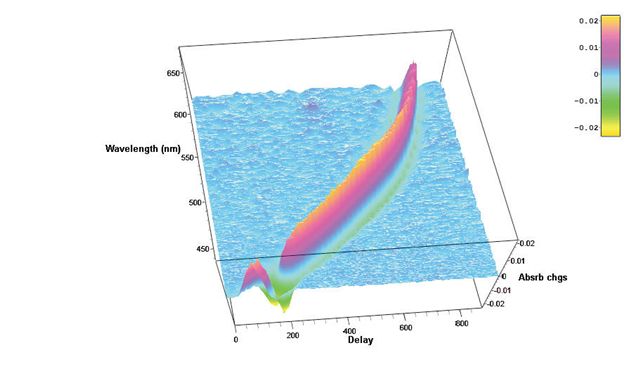 |
Hatteras-D
femtosecond transient absorption data acquisition system
Future nanostructures and biological nanosystems will take
advantage not only of the small dimensions of the objects but of the
specific way of interaction between nano-objects. The interactions
of building blocks within these nanosystems will be studied and optimized on
the
femtosecond time scale - says Sergey Egorov, President and CEO of Del Mar
Photonics, Inc. Thus we put a lot of our efforts and resources into the
development of new Ultrafast
Dynamics Tools such as our Femtosecond Transient Absorption Measurements
system Hatteras. Whether you want to
create a new photovoltaic system that will efficiently convert photon energy
in charge separation, or build a molecular complex that will dump photon energy
into local heat to kill cancer cells, or create a new fluorescent probe for
FRET microscopy, understanding of internal dynamics on femtosecond time scale
is utterly important and requires advanced measurement techniques.Reserve a
spot in our Ultrafast Dynamics Tools
training workshop in San Diego, California.
|
 |
Beacon Femtosecond Optically Gated Fluorescence Kinetic Measurement System
-
request a quote -
pdf
Beacon together with Trestles Ti:sapphire oscillator, second and third harmonic
generators. Femtosecond optical gating (FOG) method gives best temporal
resolution in light-induced fluorescence lifetime measurements. The resolution
is determined by a temporal width of femtosecond optical gate pulse and doesn't
depend on the detector response function. Sum frequency generation (also called
upconversion) in nonlinear optical crystal is used as a gating method in the
Beacon femtosecond fluorescence kinetic measurement system. We offer
Beacon-DX for operation together with Ti: sapphire femtosecond oscillators
and Beacon-DA for operation together with femtosecond amplified pulses.
Beacon Trestles -
Beacon PHAROS
Reserve a
spot in our Ultrafast Dynamics Tools
training workshop in San Diego, California.
|
Product news and updates - Training Workshops
- Featured Customer - Other News
 |
Near Field Scanning Optical Microscope
NSOM Godwit - best spatial optical resolution using the near field scanning optical microscope
(NSOM) principle
Near field scanning optical microscope (NSOM) and atomic force microscope (AFM)
modes of operation
NSOM images with laser and lamp illumination
Commerciaand custom NSOM probes
Near field optica and luminescence images in photon counting mode
NSOM images in collection and illumination modes
Transmission and reflection NSOM configurations
20 nm optical resolution (Raleigh criteria for spatial resolution)
State-of-the-art optical microscope console: simultaneous sample and tip
observation with long working distance objectives
Femtosecond and UV excitation
True single molecule detection
High-resolution AFM imaging of DNA
Godwit-uScope data acquisition and Godwit-FemtoScan image processing software
Ambient light protection with light-tight box
|
 |
Trestles LH Ti:Sapphire
laser
Trestles LH is a new series of high quality femtosecond Ti:Sapphire
lasers for applications in scientific research, biological imaging, life
sciences and precision material processing. Trestles LH includes integrated
sealed, turn-key, cost-effective, diode-pumped
solid-state (DPSS). Trestles LH lasers offer the most attractive pricing
on the market combined with excellent performance and reliability. DPSS LH
is a state-of-the-art laser designed for today’s applications. It combines
superb performance and tremendous value for today’s market and has
numerous advantages over all other DPSS lasers suitable for Ti:Sapphire
pumping. Trestles LH can be customized to fit customer requirements and
budget.
Trestles LH plus OPO
(Optical Parametric Oscillator)
Reserve a
spot in our Femtosecond lasers training
workshop in San Diego, California. Come to learn how to build a
femtosecond laser from a kit
|
 |
DPSS DMPLH lasers
DPSS DMP LH series lasers will pump your Ti:Sapphire laser.
There are LH series lasers installed all over the world pumping all makes & models
of oscillator. Anywhere from CEP-stabilized femtosecond Ti:Sapphire oscillators
to ultra-narrow-linewidth CW Ti:Sapphire oscillators. With up to 10 Watts CW
average power at 532nm in a TEMoo spatial mode, LH series
lasers has quickly proven itself
as the perfect DPSS pump laser for all types of Ti:Sapphire or dye laser.
Ideal for pumping of:
Trestles LH
Ti:Sapphire laser -
ULN ultra low-noise option
T&D-scan laser
spectrometer based on narrow line CW Ti:Sapphire laser
|
 |
Pismo pulse picker
The Pismo pulse picker systems is as a pulse gating system that lets single
pulses or group of subsequent pulses from a femtosecond or picosecond pulse
train pass through the system, and stops other radiation. The system is
perfectly suitable for most commercial femtosecond oscillators and
amplifiers.
The system can pick either single pulses, shoot bursts (patterns of single
pulses) or pick group of subsequent pulses (wider square-shaped HV pulse
modification). HV pulse duration (i.e. gate open time) is 10 ns in the default
Pismo 8/1 model, but can be customized from 3 to 1250 ns upon request or made
variable. The frequency of the picked pulses starts with single shot to 1 kHz
for the basic model, and goes up to 100 kHz for the most advanced one.
The Pockels cell is supplied with a control unit that is capable of synching
to the optical pulse train via a built-in photodetector unit, while electric
trigger signal is also accepted. Two additional delay channels are available
for synching of other equipment to the pulse picker operation. Moreover, USB
connectivity and LabView-compatible drivers save a great deal of your time
on storing and recalling presets, and setting up some automated experimental
setups. One control unit is capable of driving of up to 3 Pockels cells, and
this comes handy in complex setups or contrast-improving schemes. The system
can also be modified to supply two HV pulses to one Pockels cell unit,
making it a 2-channel pulse picker system. This may be essential for
injection/ejection purposes when building a regenerative or multipass
amplifier system.
|
 |
Tourmaline Yb-SS-1058/100 Femtosecond solid state laser system
The Yb-doped Tourmaline Yb-SS laser radiates at 1058±2 nm
with more than 1 W of average power, and enables the user to enjoy
Ti:Sapphire level power at over-micron wavelengths. This new design from Del
Mar's engineers features an integrated pump diode module for greater system
stability and turn-key operation. The solid bulk body of the laser ensures
maximum rigidity, while self-starting design provides for easy
"plug-and-play" operation.
Laser for second harmonic imaging in the retina
with the voltage sensitive dye FM4-64
New fiber optics and fiber laser products
Laser Source for Photodynamic Therapy -
request additional information and quote
AOM OEM Acousto-Optic Modulators for DPSS lasers, 1060 – 2100 nm -
request additional information and quote
AOM – pigtailed for pulsed fiber lasers, 1060-2100 nm -
request additional information and quote
Quasi-CW Yb fiber laser, 36 W in CW, 17 W pulsed -
request additional information and quote
Pulsed (AOM) fiber laser, 20 W, 1060 – 2100 nm -
request additional information and quote
Pulsed (pulse width/repetition rate – tunable) MOFA-type laser 10-W, 1060 nm
-
request additional information and quote
Eye-safe CW fiber laser, 25 W -
request additional information and quote
Yellow fiber Laser, 15 W -
request additional information and quote
10-GHz photonic link -
request additional information and quote
|
 |
780 nm and 1560 nm Femtosecond Fiber Laser
Buccaneer SHG
160/80 -
Buccaneer SHG 300/120
Femtosecond Fiber Laser with SH Generation
Wavelength (switchable): 780±5 nm (fixed) or 1560±10 nm (fixed)
Pulse Width (FWHM): <120 fs
Power output (switchable apertures):
>80 mW, 780 nm, TEM00, linearly polarized or
>160 mW, 1560 nm, TEM00, linearly polarized
Repetition rate: 70 MHz
Spatial mode: TEMoo
RF SYNC out: SMA connector (200-300 mV@50 ohm load)
Mode lock status: SMA connector (3.5/0 V) and LED
For applications in Amplifier systems seeding, Terahertz generation and
detection, Multi-photon microscopy, Ultrafast spectroscopy, Semiconductor device
characterization, Supercontinuum generation, Optical coherence tomography,
Telecommunications , Micromachining, Nonlinear Bioimaging (SHG imaging), THz
spectroscopy, Education
|
 |
New laser spectrometer
T&D-scan for research that
demands high resolution and high spectral
density in UV-VIS-NIR spectral domains - now available with
new pump option!
The
T&D-scan
includes
a CW ultra-wide-tunable narrow-line laser, high-precision wavelength meter,
an electronic control unit driven through USB interface as well as a
software package. Novel advanced design of the fundamental laser component
implements efficient intra-cavity frequency doubling as well as provides a
state-of-the-art combined ultra-wide-tunable Ti:Sapphire & Dye laser
capable of covering together a
super-broad spectral range between 275 and 1100 nm. Wavelength
selection components as well as the position of the non-linear crystal are
precisely tuned by a closed-loop control
system, which incorporates highly accurate wavelength meter.Reserve a
spot in our CW lasers training
workshop in San Diego, California. Come to
learn how to build a
CW
Ti:Sapphire laser from a kit
|
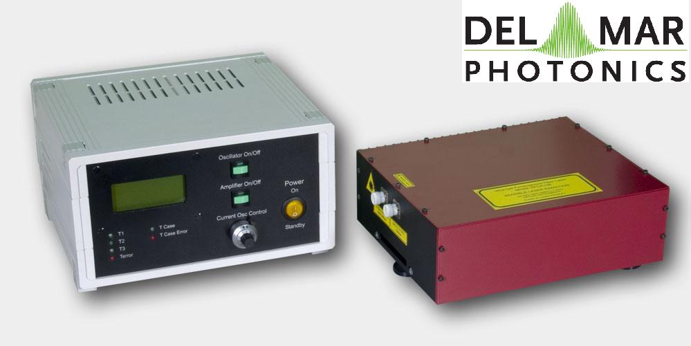
|
Femtosecond fiber laser Model Pearl-70P300
-
request a quote
Femtosecond pulsed lasers are used in many fields of physics, biology,
medicine and many other natural sciences and applications: material processing,
multiphoton microscopy, «pump-probe» spectroscopy, parametric generation and
optical frequency metrology. Femtosecond fiber lasers offer stable and steady
operation without constant realignment.
The Pearl-70P300 laser comprises: a passively mode-locked fiber laser, providing
pulses with repetition rate 60 MHz and having duration of 250-5000 fs, an
amplifier based on Er3+ doped fiber waveguide with pumping by two laser diodes,
a prism compressor for amplified pulse compression.
Pearl Ultra-Compact
Ultrafast Picosecond Fiber
Oscillator
|
|
Reef femtosecond autocorrelators
The autocorrelation technique is the most common method used to determine
laser pulse width characteristics on a femtosecond time scale.
The basic optical configuration of the autocorrelator is similar to that of an
interferometer (Figure.1). An incoming pulse train is split into two beams of
equal intensity. An adjustable optical delay is inserted into one of the arms.
The two beams are then recombined within a nonlinear material (semiconductor)
for two photon absorption (TPA). The incident pulses directly generate a
nonlinear TPA photocurrent in the semiconductor, and the detection of this
photocurrent as a function of interferometer optical delay between the
interacting pulses yields the pulse autocorrelation function. The TPA process is
polarization-independent and non-phasematched, simplifying
alignment.
Reef-RT autocorrelator measures laser
pulse durations ranging from 20 femtoseconds to picosecond regime. It measures
pulse widths from both low energy, high repetition rate oscillators and high
energy, low repetition rate amplifiers. Compact control unit operates
autocorrelator head and optional spectrometer through on-screen menus.
Autocorrelation trace and spectrum can be displayed and analyzed on screen or
downloaded to remote computer.
New:
Reef-20DDR autocorrelator
-
Multishot-FROG for femtosecond fiber laser oscillator and amplifier
Collinear (interferometric) autocorrelation
for 1300-2000 nm wavelength range |
 |
Trestles Fourth Harmonic Generator -
request a quote
Trestles FHG is designed to work with
Ti:sapphire lasers, such as the Trestles Ti:sapphire laser to provide fourth
harmonic generation. Trestles FHG is an affordable and easy solution to
generating pulses around 200 nm.
Trestles FHG is available in two versions. The first version uses the third
harmonic mixed with the fundamental wavelength to produce fourth harmonic
generation. This option provides the fundamental, as well as the second, third
and fourth harmonics. For input of 810 nm, 1 W, 82MHz and 50 fs, output of the
fourth harmonic is at 203 nm, 300-400 fs and power is 3 mW. The second option
uses two separate second harmonic generation stages to produce fourth harmonic
generation. With input of 810 nm, 1 W, 82 MHz and 50 fs, output of the fourth
harmonic is at 210 nm, pulse width of 500 fs and power of 10 mW.
The Trestles FHG from Del Mar Photonics is an easily installed solution for
adding functionality to any femtosecond laser system. Extended ranges will
increase capabilities for research, and specific applications such as microscopy
and spectroscopy.
|
 |
Near IR viewers
High performance infrared
monocular viewers are designed to observe radiation emitted by
infrared sources. They can be used to observe indirect radiation of IR
LED's and diode lasers, Nd:YAG, Ti:Sapphire, Cr:Forsterite, dye lasers and
other laser sources. IR viewers are ideal for applications involving the
alignment of infrared laser beams and of optical components in
near-infrared systems. Near IR viewers
sensitive to laser radiation up to 2000 nm.
The light weight, compact monocular may be used as a hand-held or facemask
mounted for hands free operation.
Ultraviolet viewers are
designed to observe radiation emitted by UV sources. |
 |
Hummingbird EMCCD camera
The digital Hummingbird
EMCCD camera combines high sensitivity, speed and high resolution.
It uses Texas Instruments' 1MegaPixel Frame Transfer Impactron device which
provides QE up to 65%.
Hummingbird comes with a standard CameraLink output.
It is the smallest and most rugged 1MP EMCCD camera in the world.
It is ideally suited for any low imaging application such as hyperspectral
imaging, X-ray imaging, Astronomy and low light surveillance.
It is small, lightweight, low power and is therefore the ideal camera for
OEM and integrators.
buy online |
Training Workshops
Featured Customer
 |
Trestles LH10-fs/CW laser system at UC Santa Cruz Center of
Nanoscale Optofluidics
Del
Mar Photonics offers new
Trestles fs/CW laser system which can be easily
switched from femtosecond mode to CW and back. Having both modes of operation in one system dramatically increase a
number of applications that the laser can be used for, and makes it an ideal
tool for scientific lab involved in multiple research projects.
Kaelyn Leake is a PhD student in Electrical Engineering. She graduated from
Sweet Briar College with a B.S. in Engineering Sciences and Physics. Her
research interests include development of nanoscale optofluidic devices and
their applications. Kaelyn is the recipient of a first-year QB3 Fellowship.
In this video Kaelyn talks about her experimental research in nanoscale
optofluidics to be done with Trestles LH laser.
Reserve a spot in our
femtosecond Ti:Sapphire training workshop in San
Diego, California during summer 2011 |
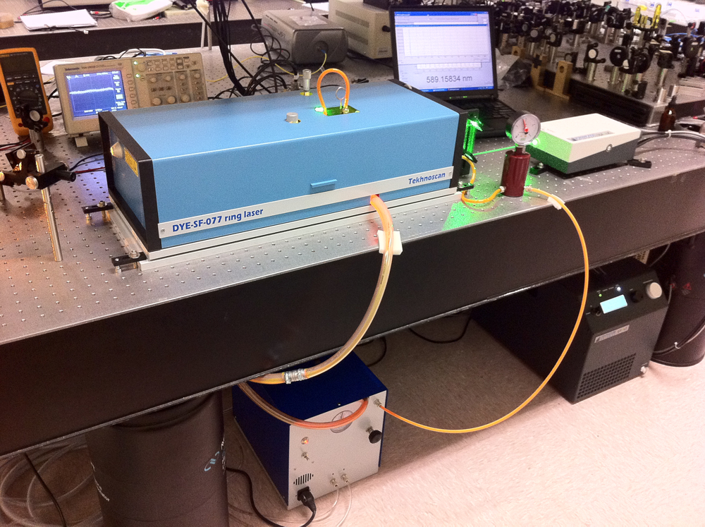 |
Frequency-stabilized CW
single-frequency ring Dye laser DYE-SF-077 pumped by DPSS DMPLH laser installed
in the brand new group of Dr. Dajun Wang at the The Chinese University of Hong
Kong.
DYE-SF-077 features exceptionally narrow generation line width, which
amounts to less than 100 kHz. DYE-SF-077 sets new standard for generation
line width of commercial lasers. Prior to this model, the narrowest line-width
of commercial dye lasers was as broad as 500 kHz - 1 MHz. It is necessary to
note that the 100-kHz line-width is achieved in DYE-SF-077 without the use of an
acousto-optical modulator, which, as a rule, complicates the design and
introduces additional losses. A specially designed ultra-fast PZT is used for
efficient suppression of radiation frequency fluctuations in a broad frequency
range. DYE-SF-077 will be used in resaerch of Ultracold polar molecules,
Bose-Einstein condensate and quantum degenerate Fermi gas and High resolution
spectroscopy
630nm |
Del Mar Photonics continuously expands its components
portfolio.
 |
We are looking forward to hear from you and help you with
your optical and crystal components requirements. Need time to think about
it?
Drop us a line and we'll send you beautiful Del Mar Photonics mug (or
two) so you can have a tea party with your colleagues and discuss your
potential needs. |
Del Mar Photonics, Inc.
4119 Twilight Ridge
San Diego, CA 92130
tel: (858) 876-3133
fax: (858) 630-2376
Skype: delmarphotonics
sales@dmphotonics.com